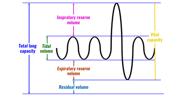
36 label the respiratory volumes on the diagram using the terms provided.
Lung volume is the volume of gas during each phase/ part of respiration. For example, there is a certain volume of gas we all inhale during our normal respiration (i.e respiration during rest). Now, when I ask you to inhale forcefully to the maximum capacity, we take the extra volume of air inside. Using the terms provided to match up with the description concerning respiratory volume measurements: A. Dead space volume B. Expiratory reserve volume (ERV) C. Tidal volume (TV) D. Residual volume (RV) E. Inspiratory reserve volume (IRV) F. Vital capacity (VC) _____ i) Respiratory volume inhaled or exhaled during normal breathing _____ ii) Air ...
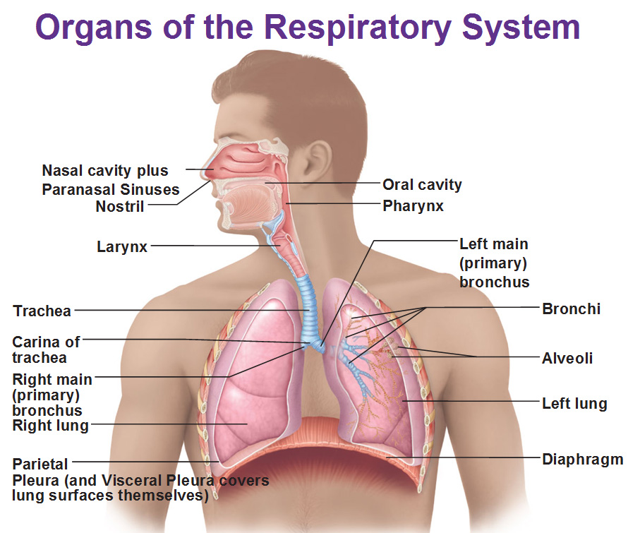
It is useful to divide the total space within the lungs into volumes and capacities. These can be measured to aid in the definitive diagnosis, quantification and monitoring of disease. They allow an assessment of the mechanical condition of the lungs, its musculature, airway resistance and the effectiveness of gas exchange at the alveolar membrane while being, for the most part, cheap, non ...

Label the respiratory volumes on the diagram using the terms provided.
Label the respiratory capacities on the diagram using the terms provided. Image: Label the respiratory capacities on the diagram using the terms provided. Lung volumes measurement is an integral part of pulmonary function test. These volumes tend to vary, depending on the depth of respiration, ethnicity, gender, age, body composition and in certain respiratory diseases. A number of the lung volumes can be measured by Spirometry- Tidal volume, Inspiratory reserve volume, and Expiratory reserve volume. 1. Using the information provided in Table 3-1 and Figure 3-1, what are the values for tidal volume, inspiratory capacity, expiratory reserve volume, functional residual capacity, vital capacity, and total lung capacity? (Hint: It may be helpful to label the spirometry diagram with the names of the lung volumes and capacities.) 2.
Label the respiratory volumes on the diagram using the terms provided.. Regions of the Digestive System. At its simplest, the digestive system is a tube running from mouth to anus.Its chief goal is to break down huge macromolecules (proteins, fats and starch), which cannot be absorbed intact, into smaller molecules (amino acids, fatty acids and glucose) that can be absorbed across the wall of the tube, and into the circulatory system for dissemination throughout ... Introduction. Severe illness characterised by progressive hypoxaemic respiratory failure develops in a large number of patients with COVID-19, resulting in the need for invasive mechanical ventilation.1, 2, 3 In patients who are intubated and have moderate to severe acute respiratory distress syndrome, prone positioning is an effective intervention to improve oxygenation and reduce mortality.4 ... The diagram below shows the structure and functions of the human digestive system. Let learn the different parts of the human digestive system. Mouth — It includes teeth, salivary glands and tongue. It is the beginning of the digestive tract and the process of digestion begins from the mouth, where teeth help by breaking and grinding the food ... Label the anterior view of the larynx based on the hints if provided. alveolar duct. ... label the respiratory muscles and the respiratory centers that control them. epiglottis. ... These symptoms lead to temporary changes in respiratory air volumes and capacities.
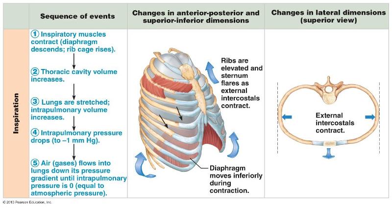
Body Cavities. The cavities, or spaces, of the body contain the internal organs, or viscera.The two main cavities are called the ventral and dorsal cavities. The ventral is the larger cavity and is subdivided into two parts (thoracic and abdominopelvic cavities) by the diaphragm, a dome-shaped respiratory muscle.. Thoracic cavity Waves move freely and label the diagram respiratory system is the digestive system and complete the skeleton diagram muscular system and this question submission via the perfect. Are labels for diagrams and skull diagram worksheet answers, expand your child working on either cause neighboring neurons appear to. The best way to upload files is by using the "additional materials" box. Drop all the files you want your writer to use in processing your order. If you forget to attach the files when filling the order form, you can upload them by clicking on the "files" button on your personal order page. 2. Use the provided values to construct lung volume graphs on graph paper of lung volume (mL) vs time. Show multiple cycles of tidal breathing and at least 1 maximum exhalation and 1 maximum inhalation. Label all the different lung volumes using arrows and labels. It may help to first determine the missing lung volumes of each data set. a.
Showing top 8 worksheets in the category integumentary system grade 6. Worksheet the integumentary system answer key label the diagram in the spaces provided. Vitamin d synthesis begins in skin exposed to uv light. Functions of the skin skin is a barrier to microbes chemical irritants water loss. Start studying integumentary system 6th grade. Identify the anatomical structures of the upper respiratory tract. Label the respiratory capacities on the diagram using the terms provided. Classify the definition or example with the correct characteristic of sound. Classify the airflow factors on whether they would have an effect on resistance or pressure gradient. Label the respiratory volumes on the diagram using the terms provided. Label the respiratory capacities on the diagram using the terms provided. Put the processes of gas exchange into the correct order, beginning with inhalation. Complete each statement using a term or terms from the list below. Write your answers in the spaces provided. Some words may be used more than once. heart circulation oxygen veins food capillaries arteries blood vessel waste materials blood 1. The transport of materials in living things is called _____. 2.
Closeup of skeleton hand model
four. The volume of air that enters and leaves the lungs can be measured with an instrument called a _______. spirometer. Fill in the blanks with the appropriate terms regarding the general functions of the respiratory system. respiratory system facts. Oxygen bound to hemoglobin is referred to as _______. Oxyhemoglobin.

35 Label The Respiratory System Answers - Labels For You
Transcribed image text: 1. Using the terms from the table above to label the parts in this image (a-h). 2. Identify the various lung volumes and capacities in the following diagram: Forced inspiration Quy aunon Bung breathing -Forced ton -Volume of all Table 27-1 Respiratory Volumes and Capacities Volumes and Capacities Definition Tidal volume (TV) Volume of air exchanged during each normal ...
The Respiratory System
Label the respiratory capacities on the diagram using the terms provided. Image: Label the respiratory capacities on the diagram using the terms provided.
Respiratory Volumes & Lung Capacity Explained - TeachPE.com
Label the respiratory volumes on the diagram below, using these terms. expiratory reserve volume inspiratory reserve volume > residual volume > tidal volume total lung capacity > vital capacity F Win E A = B = = C= D E = F =
Solved: PART G ASSESSMENTS Label The Major Arteries And Ve ...
Start studying QUIZ Chapter 18 The respiratory system. Learn vocabulary, terms, and more with flashcards, games, and other study tools. Search. Browse. Create. ... Tidal volume (TV) ... Label the diagram using the lung columes and capacities you listed above.
Longitudinal changes in clinical outcomes and candidate ...
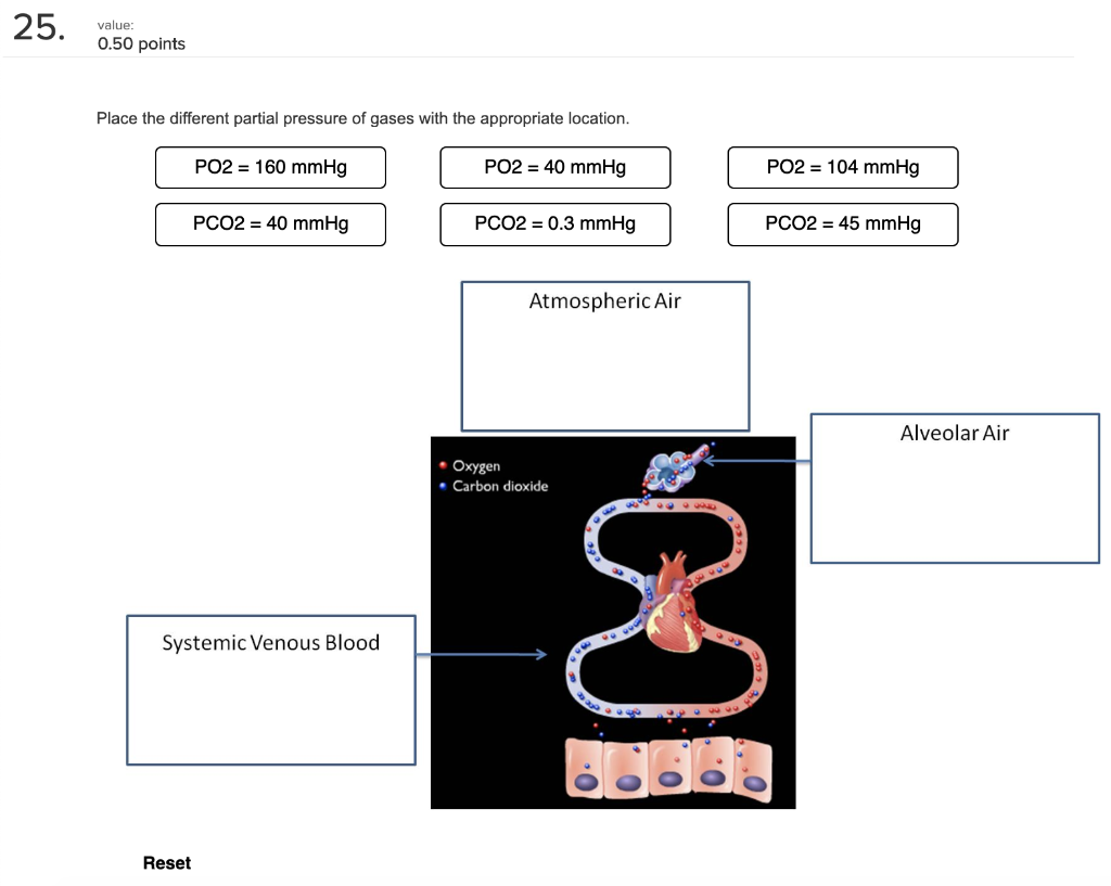
Fill in the blanks with the terms provided. Not all labels will be used. The P(O₂) in the alveoli is 104 mm Hg, and the blood ...
![ibbio [licensed for non-commercial use only] / Gas exchange](http://ibbio.pbworks.com/f/1305621432/alveolus-of-man.jpg)
ibbio [licensed for non-commercial use only] / Gas exchange
Oct 31, 2018 · Paranasal sinuses conveys air from larynx to bronchial tree. Label the respiratory volumes on the diagram using the terms provided.. Appropriately label all structures provided with leader lines on the diagrams below. It is useful to divide the total space within the lungs into volumes and capacities. Bronchials trace a molecule of oxygen from the nostrils to the pulmonary capillaries of the lunges.
30 Label The Respiratory System Answers - Labels Database 2020
Label the respiratory volumes on the diagram using the terms provided Fill in the blanks with the appropriate terms The most important stimulus affecting breathing rate and depth is BLOOD P(CO2)
Chapter 44 Solutions | Laboratory Manual For Human Anatomy ...
(Some states use other terms for "infectious medical waste," such as "regulated medical waste" or "special waste.") OSHA. 29 CFR Part 1910.1030, Occupational Exposure to Bloodborne Pathogens, provides minimal requirements for labeling and packaging of blood and body fluids when transported or outside a laboratory.
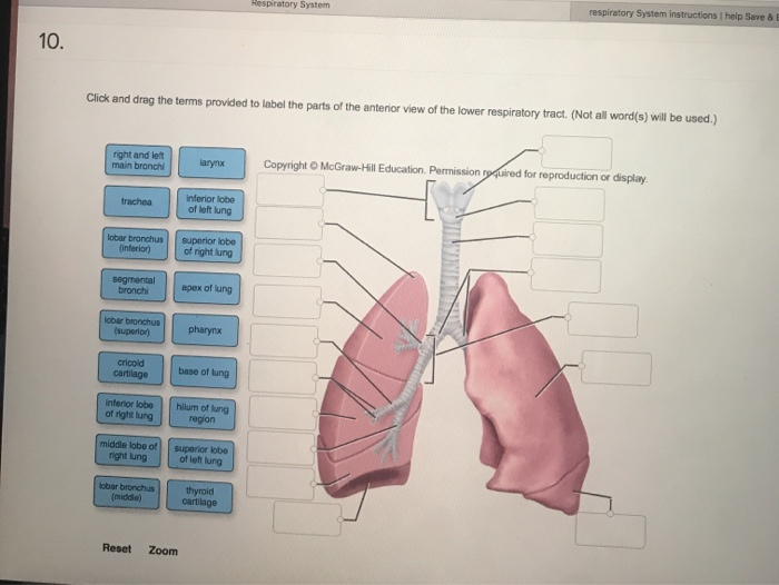
Solved: Hespiratory System Respiratory System Instructions ...
Label the diagram below using the checklist provided. Complete Nasal Cavity Structure: Epiglottis Pharynx Larynx ... Define the following respiratory terms a) b) Think about how your breathing changes during exercise. Explain what is happening to each ... Explain why the athlete's tidal volume has changed after 40 minutes of playing hockey.
Chapter 22 Flashcards | Easy Notecards
Correctly label the following diagram relating blood vessel type to total cross-sectional area and velocity of blood flow. ... Heart rate and stroke volume decrease under decreased parasympathetic stimulation. ... Using the terms provided, trace a drop of blood from the posterior side of the knee back to the heart. ...

Respiration and Circulation - Mrs Smallwood's Science Site
The following terms describe the various lung (respiratory) volumes: The tidal volume (TV), about 500 mL, is the amount of air inspired during normal, relaxed breathing.; The inspiratory reserve volume (IRV), about 3,100 mL, is the additional air that can be forcibly inhaled after the inspiration of a normal tidal volume.; The expiratory reserve volume (ERV), about 1,200 mL, is the additional ...

Solved: Label The Tissues And Structures On This Histology ...
Study design . A prospective, randomized, single-blind, 2-arm, parallel-group, open-label, phase I-II clinical trial was performed between May and December 2020 and carried out according to the consolidated standards of reporting trials (CONSORT) statement guidelines for randomized controlled trials ().The study aimed to evaluate the safety and efficacy of electrolyzed saline for preventing ...
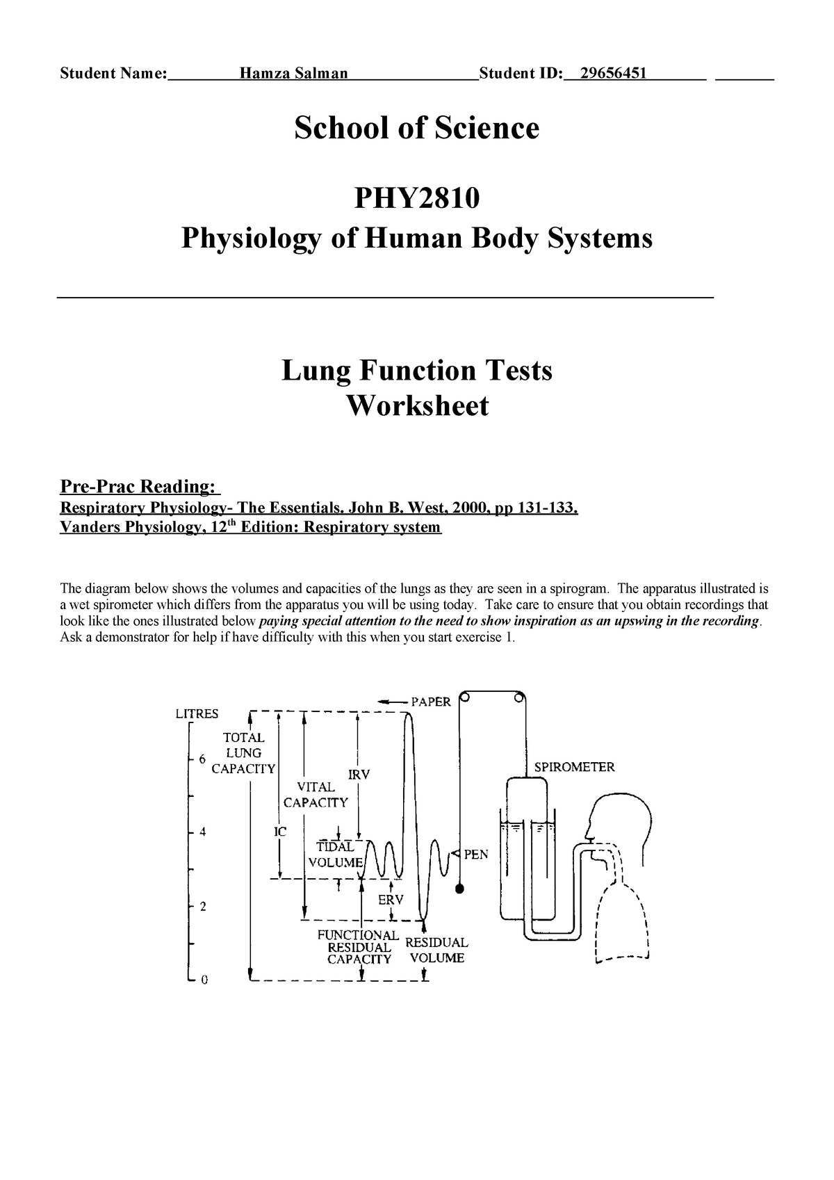
Lung Function Worksheet - PHY2810 - StuDocu
You will be able to describe the body's regions using the terms from the figure. Directional Terms. Certain directional anatomical terms appear throughout this and any other anatomy textbook (Figure 1.4.2). These terms are essential for describing the relative locations of different body structures.
33 Label The Respiratory System Worksheet - Labels For You
Anatomy and Physiology is a dynamic textbook for the yearlong Human Anatomy and Physiology course taught at most two- and four-year colleges and universities to students majoring in nursing and allied health. A&P is 29 chapters of pedagogically effective learning content, organized by body system, and written at an audience-appropriate level.
BIO 161 Worksheet 8 - BIO 161 - Cal Poly - StuDocu
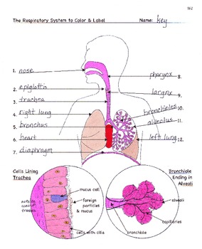
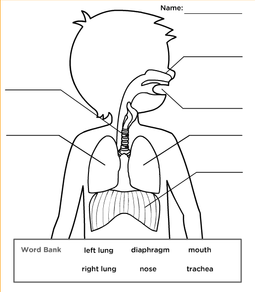
ACTIVITY - Label the Respiratory System Name: _____ Date: _____ Block: _____ Label the diagram of the respiratory system below with the following parts, then colour your diagram. left bronchus trachea mouth pharynx (throat) diaphragm

Closeup of skeleton hand model
Residual Volume (RV): Volume of air r emaining in the lungs even after a forcible expiration. This averages 1100 mL to 1200 mL. By adding up a few respiratory volumes described above, one can derive various pulmonary capacities, which can be used in clinical diagnosis. Inspiratory Capacity (IC): Total volume of air a person can inspire

Lymphatic System Labeled Diagram Stock Illustration ...
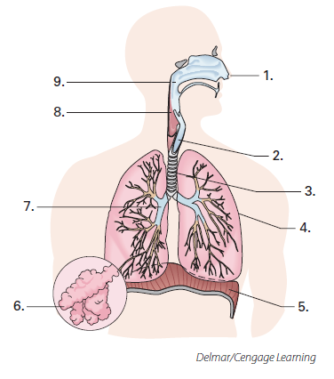
The respiratory system helps in breathing (also known as pulmonary ventilation.) The air inhaled through the nose moves through the pharynx, larynx, trachea and into the lungs. The air is exhaled back through the same pathway. Changes in the volume and pressure in the lungs aid in pulmonary ventilation. Exchange of Gases between Lungs and ...

32 Label The Structures Of The Bone Using The Hints ...
The respiratory system is the organs and other parts of your body involved in breathing when you exchange oxygen and carbon dioxide. Drag the labels onto the diagram to identify respiratory system structures. It is made up of several organs and structures that transport air into and out of the lungs exchanging oxygen with carbon dioxide.

A&P CH22 The Respiratory System Flashcards | Easy Notecards
Capillaries - Structure & Function Explained with Diagrams. Capillaries are the smallest of all blood vessels and form the connection between veins and arteries. As arteries branch and divide into arterioles and continue to reduce in size as they reach the muscle they become capillaries. Here the capillaries form a capillary bed, which is a ...
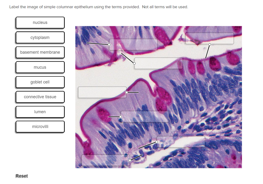
Solved: Label The Image Of Simple Columnar Epithelium Usin ...
2 32 Chapter 13 The Respiratory System 15. The following section concerns respiratory volume measurements. Using key choices, select the terms identified in the following descriptions by inserting the appropriate term or letter in the answer blanks. Key Choices A. Dead space volume C. Inspiratory reserve volume (IRV) E. Tidal volume (TV)
Solved: 23. Value: 0.50 Points Label The Anterior View Of ...
Expiratory reserve volume Quantity of air that can be forcefully expired after a normal tidal expiration. 1200mL Vital capacity Maximum quantity of air that can be moved into and out of the lungs VC = IRV+ERV+TV 4500-5500mL Functional residual capacity Volume of lung after normal exhalation FRC = ERV+RV 2400mL
33 Label The Structures Of The Respiratory System - Labels ...
1. Using the information provided in Table 3-1 and Figure 3-1, what are the values for tidal volume, inspiratory capacity, expiratory reserve volume, functional residual capacity, vital capacity, and total lung capacity? (Hint: It may be helpful to label the spirometry diagram with the names of the lung volumes and capacities.) 2.
30 Label The Respiratory System Answers - Labels Database 2020
Lung volumes measurement is an integral part of pulmonary function test. These volumes tend to vary, depending on the depth of respiration, ethnicity, gender, age, body composition and in certain respiratory diseases. A number of the lung volumes can be measured by Spirometry- Tidal volume, Inspiratory reserve volume, and Expiratory reserve volume.
Segmentation analysis of the subsolid nodule demonstrated ...
Label the respiratory capacities on the diagram using the terms provided. Image: Label the respiratory capacities on the diagram using the terms provided.

IB SEHS 2.1 Ventilatory System Flashcards | Quizlet
Solved: Uaits Conversion-The Factor Label Method Page B-4 ...
A&P CH22 The Respiratory System Flashcards | Easy Notecards
FIGURE 3.21. Microtubules are responsible for the movement ...

Free Anatomy Quiz - The Respiratory System Image

Computed tomography (CT) tumor volume measurement in a 73 ...
Solved: 8. Appropriately Label All Structures Provided Wit ...

33 Label Respiratory System Answers - Labels Information List
Closeup of skeleton pelvic model

Turkish Cement Supplier | Kdz. Eregli sea port

CyberSurgeons
0 Response to "36 label the respiratory volumes on the diagram using the terms provided."
Post a Comment