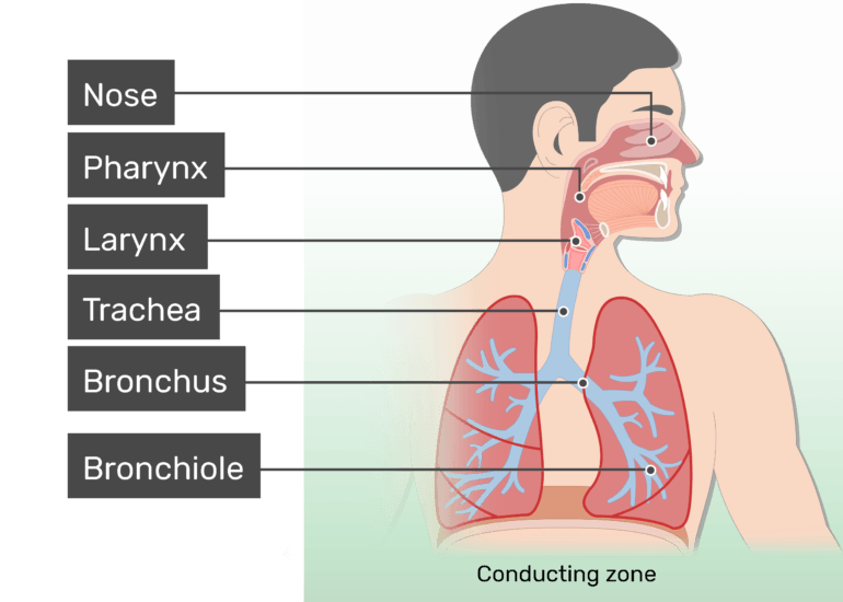
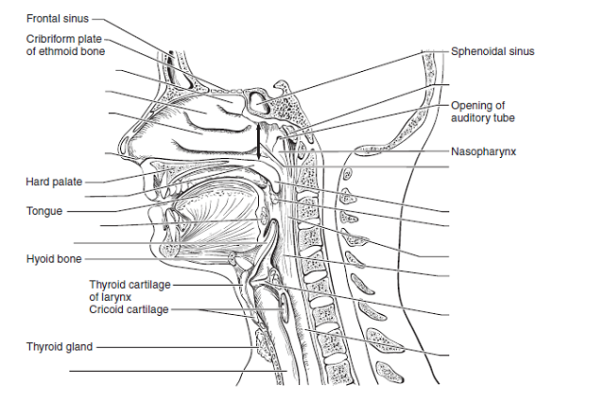
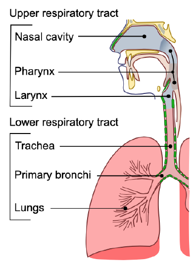
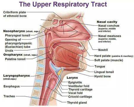
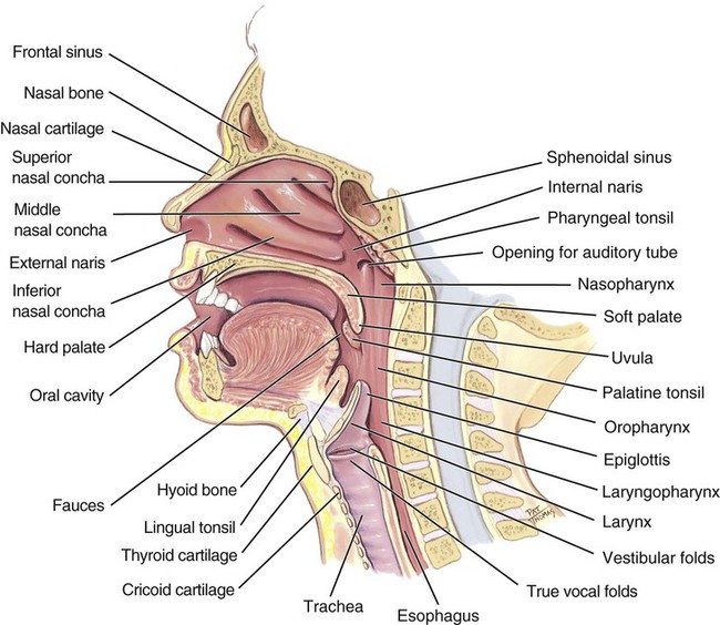
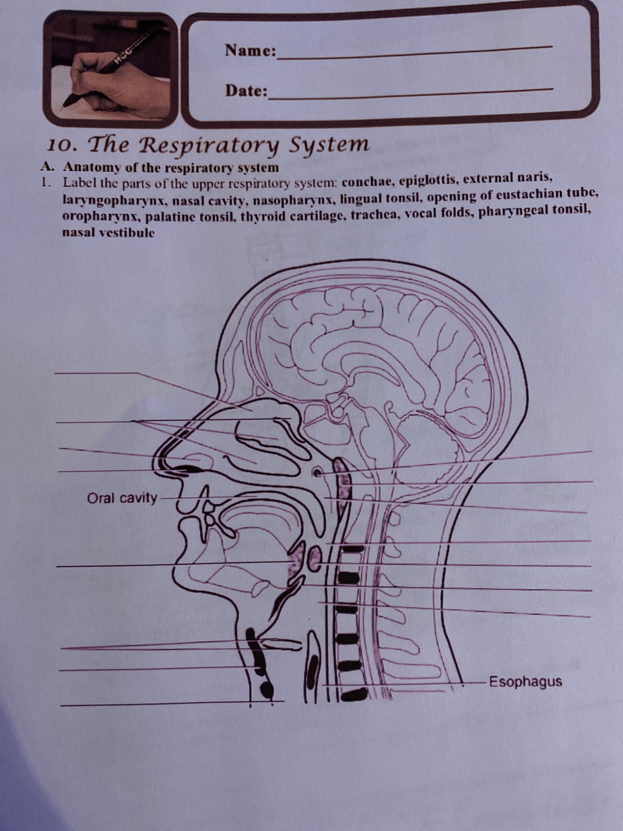
35 complete the labeling of the diagram of the upper respiratory structures
Placenta previa: This condition occurs when the placenta forms partially or totally toward the lower end of the uterus, including the cervix, rather than closer to its upper part. In cases of complete previa, the internal os—that is, the opening from the uterus to the vagina—is completely covered by the placenta. Occurring in about 1 in 200 ... Anatomy is the science that studies the structure of the body. On this page, you'll find links to descriptions and pictures of the human body's parts and organ systems from head to toe.
The anatomy of the human face is comprised of more bones than just one skull--there are 14 bones, all with different functions. Explore the different facial bones, where they are in the human face ...

Complete the labeling of the diagram of the upper respiratory structures
The head and neck, as a general anatomic region, is characterized by a large number of critical structures situated in a relatively small geographic area. It is inclusive of osseous, nervous, arterial, venous, muscular, and lymphatic structures. Lymphadenopathy is a significant clinical finding associated with acute infection, granulomatous disease, autoimmune disease, and malignancy. One of the first things you will notice if you look at the 12 steps is the pattern between the right and left side of the heart is similar. Step 1 and 6 involve a blood vessel, which makes sense as this is how blood enters and exits that side of the heart. Steps 2-5 involve a chamber, valve, chamber, and valve. While the primary function of the respiratory system is gas exchange, this extensive organ system also has some other roles. In humans and other mammals, the respiratory system is integral creating sounds such as those used for speech. Structures of the upper respiratory tract, especially the larynx, are involved in the production of sound and can modulate pitch, volume, and clarity.
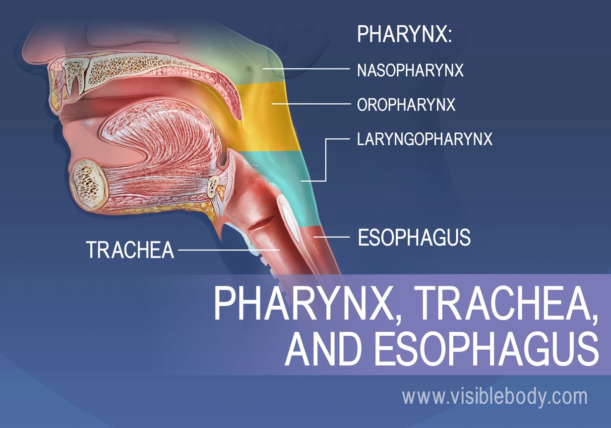
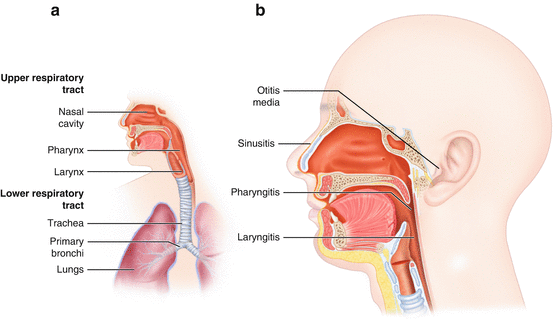
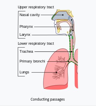
Complete the labeling of the diagram of the upper respiratory structures. As you can see in the diagram, the four abdominal quadrants are located in the space directly below the diaphragm. ... Definition and Structures 6:14 ... Diseases of the Upper Respiratory System ... Taste buds are a small organ located primarily on the tongue. The adult human tongue contains between 2,000 and 8,000 taste buds, each of which are made up of 50 to 150 taste receptor cells. Taste receptor cells are responsible for reporting the sense of taste to the brain . It used to be believed that the tongue was divided like a map into ... Anatomy of Respiratory System: Organs and Functions. The three major parts of the respiratory system all work together to carry out their task. The airways (nose, mouth, pharynx, larynx etc.) allow air to enter the body and into the lungs. The lungs work to pass oxygen into the body, whilst removing carbon dioxide from the body. The inside of the nose, including the bones, cartilage and other tissue, blood vessels and nerves, all the way back posteriorly to the nasopharynx, is called the nasal cavity. It is considered part of the upper respiratory tract due to its involvement in both inspiration and exhalation.
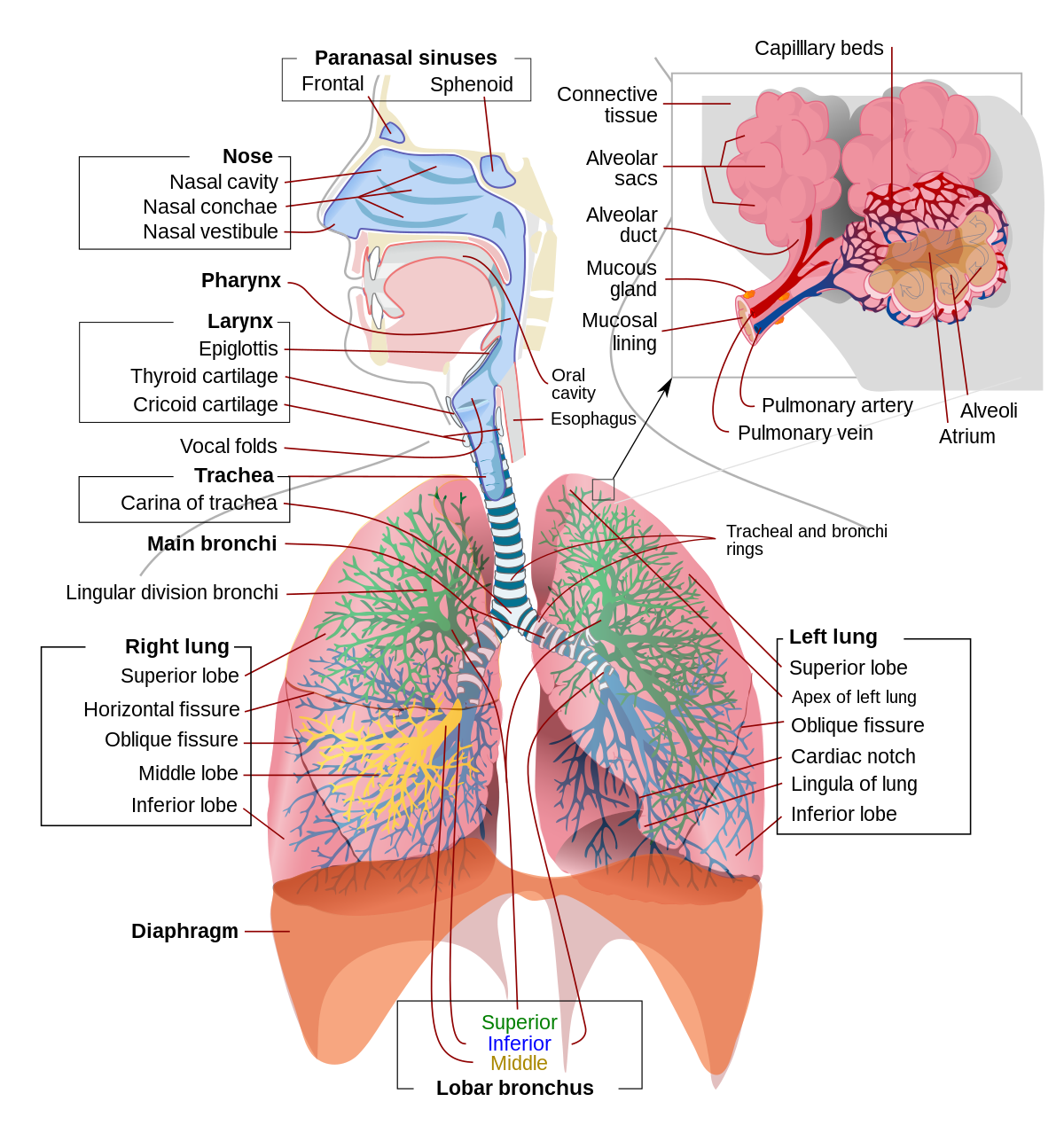
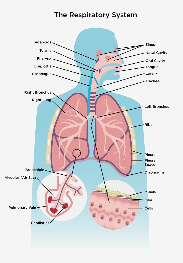
The muscles in the anterior compartment of the forearm are organised into three layers:. Superficial: flexor carpi ulnaris, palmaris longus, flexor carpi radialis, pronator teres.; Intermediate: flexor digitorum superficialis.; Deep: flexor pollicis longus, flexor digitorum profundus and pronator quadratus.; This muscle group is associated with pronation of the forearm, flexion of the wrist ... Author's Note: I made this as a kinda Wikipedia style biological summary of Valfalk/Valphalk/Valstrax with speculative features and explanations for its abilities. Do note that this is heavily speculative and mostly based on my very basic understanding of biology and general observation with these monsters. It is all labelled so you can skip to specific parts if you want. However, I put a lot of effort into this one in particular (Almost double the word count on the Zinogre post) and I’d recomme... Take a look at the labeled diagram of the respiratory system above. As you can see, there are several structures to learn. Spend a few minutes reviewing the name and location of each one, then try testing your knowledge by filling in your own diagram of the respiratory system (unlabeled) using the PDF download below. Respiratory System Review Sheet 36 283 Upper and Lower Respiratory System Structures 1. It includes the nose mouth larynx trachea bronchial tubes lungs diaphragm and muscles that enable breathing. Complete the labeling of the diagram of the upper respiratory structures sagittal section.
Discover the structures that make up the upper and lower limbs, how the limbs are connected to the axial skeleton, and the importance of the appendicular skeleton. Updated: 08/24/2021 Create an ... This is the term used to describe the tree-like structure of passageways that brings air into the lungs. The walls of the alveoli are very thin. This lets oxygen and CO2 pass easily between the alveoli and capillaries, which are very small blood vessels. One cubic millimeter of lung tissue contains around 170 alveoli. Important Biology diagrams for CBSE Class 10 are given below: 1. Neuron. Neurons or the nerve cells form the basic components of the nervous system. A typical neuron possesses a cell body called ... Nervous system practice test. First things first: you're going to need to get in some practice. After some initial reading around the subject (we recommend our article on The Nervous System), one of our favorite ways to get acquainted with a new anatomical topic is with a labeling exercise.Think of it as a sort of nervous system practice test run.
The thorax is the region between the abdomen inferiorly and the root of the neck superiorly.[1][2] It forms from the thoracic wall, its superficial structures (breast, muscles, and skin) and the thoracic cavity.
The paranasal sinuses are air-filled extensions of the nasal cavity. There are four paired sinuses - named according to the bone in which they are located - maxillary, frontal, sphenoid and ethmoid. Each sinus is lined by a ciliated pseudostratified epithelium, interspersed with mucus-secreting goblet cells.
Respiratory System SHEET Upper and Lower Respiratory System Structures 1. Complete the labeling of the model of the respiratory structures (sagittal section) shown below. Nasal "Conchal Nasal meatus REVIEW as a distibule Hard plate -posterior nasal aperature -s of t palate "uvula -palentine tonsil epiglottis vestibular fold vocal fold Thyroid ...
Inside Out Anatomy the Respiratory System - This black and white diagram is another excellent worksheet for labeling parts of the respiratory system. Lungs Anatomy Worksheet - This fun worksheet not only has kids label the parts of the respiratory system but then use the labels to solve a crossword puzzle.
The respiratory system provides oxygen to the body's cells while removing carbon dioxide, a waste product that can be lethal if allowed to accumulate. There are 3 major parts of the respiratory system: the airway, the lungs, and the muscles of respiration.
human skeleton labelled images. 10,477 human skeleton labelled stock photos, vectors, and illustrations are available royalty-free. See human skeleton labelled stock video clips. of 105. skeletal system anatomy labelled skeleton skeleton label skeletal system posterior view labeled skeleton skeleton labeled skeleton labels male human skeletle ...
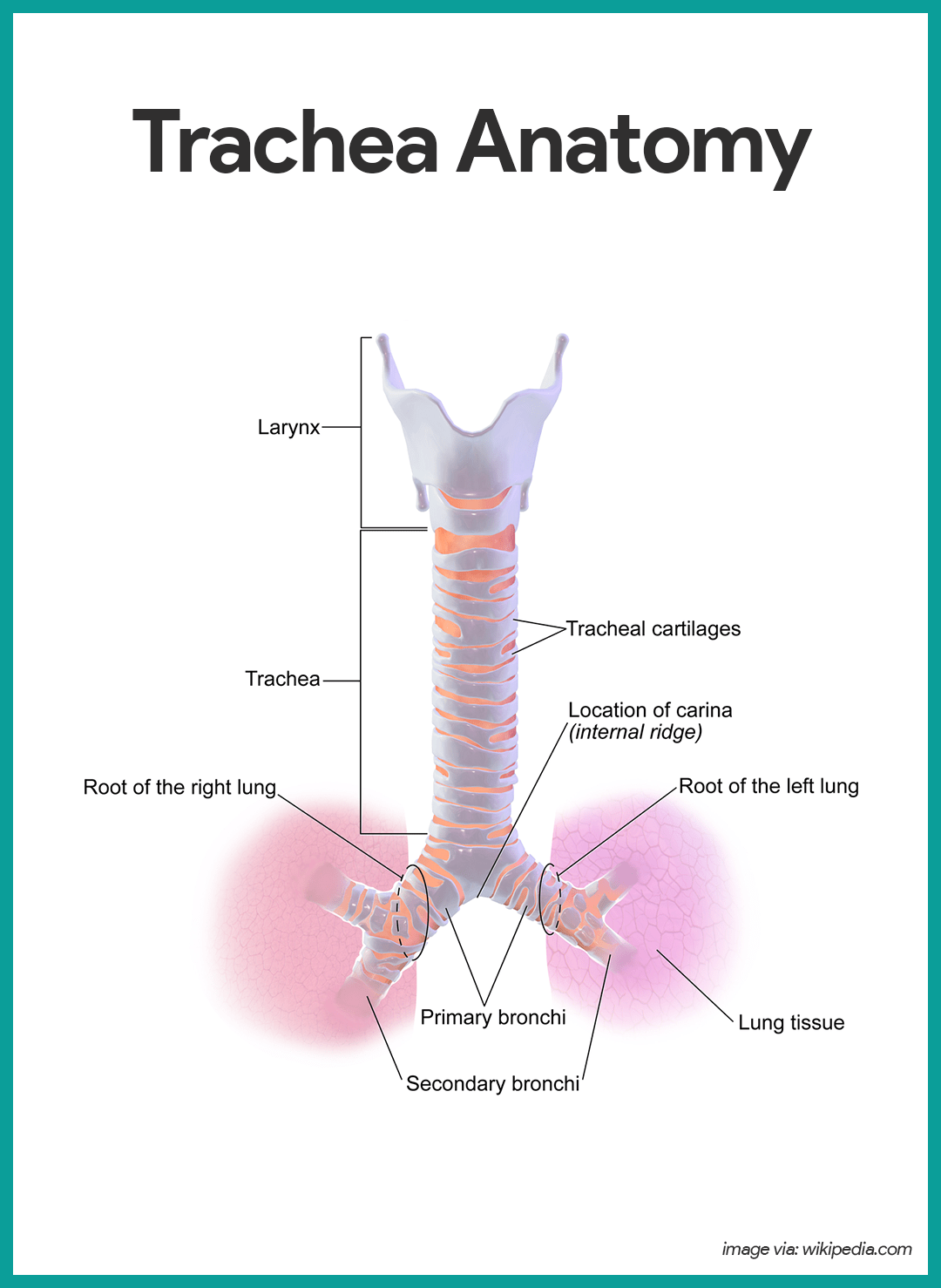
The trachea, or windpipe, is a 10-11 cm long fibrocartilaginous tube of the lower respiratory tract.It forms the trunk of the tracheobronchial tree, or pulmonary conducting zone.The trachea extends between the larynx and thorax, consisting of two parts; cervical and thoracic.It ends at the level of the sternal angle (T5) where it divides into two main bronchi, one for each lung.
The respiratory portion of the lung consists of respiratory bronchiole, alveolar duct, alveolar sac, and finally alveoli where actual respiration takes place. In the conducting zone, the air is moistened, warmed, and filtered before it reaches the start of the respiratory region at the respiratory bronchioles.
Lymphatic system (anterior view) The lymphatic system is a system of specialized vessels and organs whose main function is to return the lymph from the tissues back into the bloodstream.. Lymphatic system is considered as a part of both the circulatory and immune systems, as well as a usually neglected part of students' books. The functions of the lymphatic system complement the bloodstream ...
Anatomy and Physiology is a dynamic textbook for the yearlong Human Anatomy and Physiology course taught at most two- and four-year colleges and universities to students majoring in nursing and allied health. A&P is 29 chapters of pedagogically effective learning content, organized by body system, and written at an audience-appropriate level.
Function and anatomy of the heart made easy using labeled diagrams of cardiac structures and blood flow through the atria, ventricles, valves, aorta, pulmonary arteries veins, superior inferior vena cava, and chambers. Includes an exercise, review worksheet, quiz, and model drawing of an anterior vi
Skull The skull is a strong, bony capsule that rests on the neck and encloses the brain. It consists of two major parts: the neurocranium (cranial vault) and the viscerocranium (facial skeleton). The neurocranium is the part enveloping the brain and is formed out of two parts; the skull base that supports the brain and the calvaria (skullcap) that sits on top of the base, covering the brain.
While the primary function of the respiratory system is gas exchange, this extensive organ system also has some other roles. In humans and other mammals, the respiratory system is integral creating sounds such as those used for speech. Structures of the upper respiratory tract, especially the larynx, are involved in the production of sound and can modulate pitch, volume, and clarity.
One of the first things you will notice if you look at the 12 steps is the pattern between the right and left side of the heart is similar. Step 1 and 6 involve a blood vessel, which makes sense as this is how blood enters and exits that side of the heart. Steps 2-5 involve a chamber, valve, chamber, and valve.
The head and neck, as a general anatomic region, is characterized by a large number of critical structures situated in a relatively small geographic area. It is inclusive of osseous, nervous, arterial, venous, muscular, and lymphatic structures. Lymphadenopathy is a significant clinical finding associated with acute infection, granulomatous disease, autoimmune disease, and malignancy.










/human-respiratory-system-lungs-anatomy-953787016-b751ff559dc2489abdceb18b8fb77e8f.jpg)
0 Response to "35 complete the labeling of the diagram of the upper respiratory structures"
Post a Comment