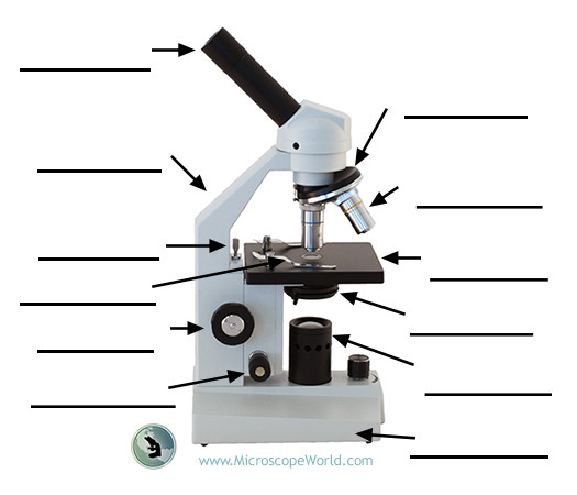
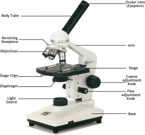
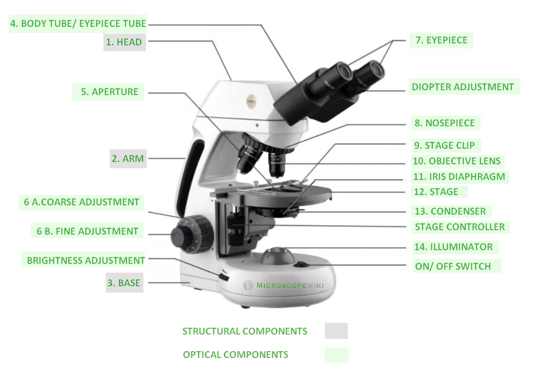
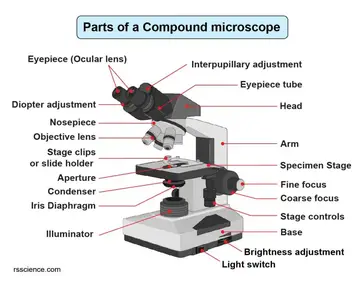
35 diagram of a compound light microscope
Compound Microscope. Definition: Compound microscopes are dual-lens microscopes. The two lenses are called objective lens and eyepiece. It is commonly referred to as a student microscope. Lens used: A compound microscope uses two convex lenses, one in the eyepiece and another in the objective. The eyepiece lenses vary with their focal lengths and hence the magnifications also differ.
**PART ONE HUNDRED AND SIXTY-FIVE** For the first two hours, Dr Kearns did most of the talking, reminding Boyd of the progress he’d made and the milestones he had achieved in his life. All the while, Boyd sat slumped forward with his forearms pressed across his knees, allowing the words of praise to wash over him, knocking back the tidal wave of doubt that had engulfed him outside. On the rare occasion that Doctor Kearns asked him for input, Boyd kept his answers brief. One sentence here and th...
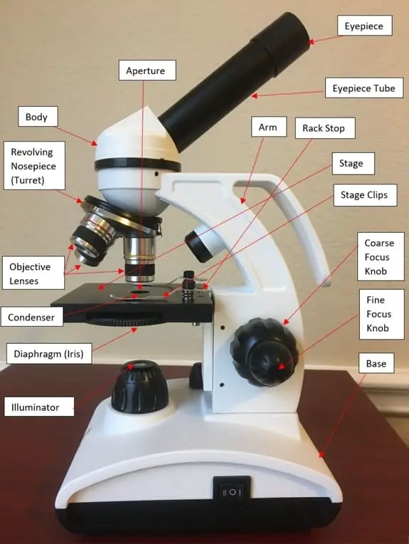
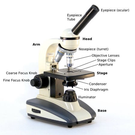
And with the help of the handy microscope diagram and microscope worksheet found on this page, you’ll be an expert on light microscope parts in no time. Together, these two science worksheets make a great study guide for students preparing for an upcoming parts of a compound microscope quiz or freshman biology test.

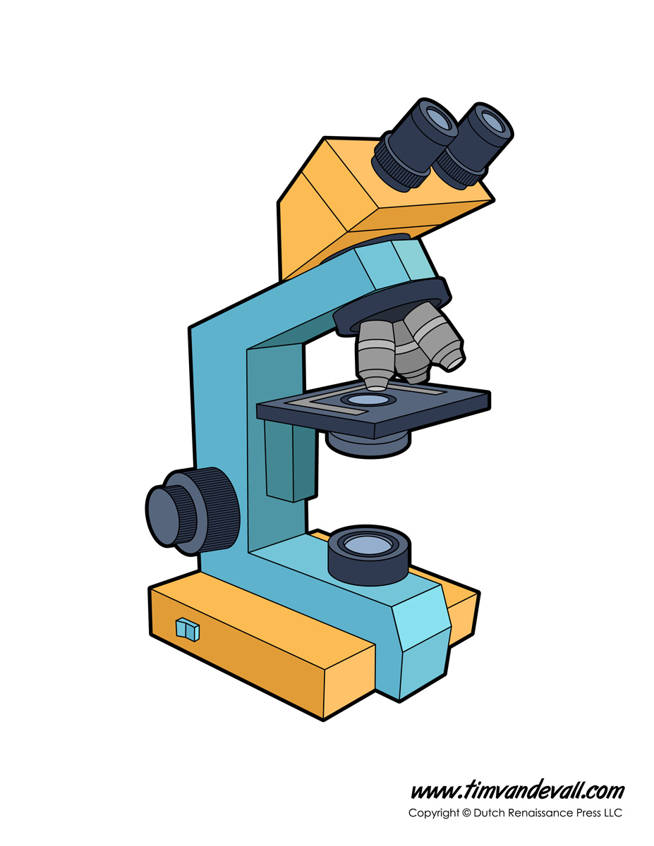
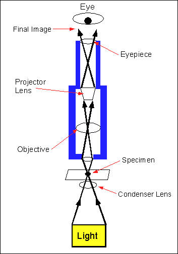
Diagram of a compound light microscope
Compound Microscope - Diagram (Parts labelled), Principle and Uses. Also called as binocular microscope or compound light microscope, it is a remarkable magnification tool that employs a combination of lenses to magnify the image of a sample that is not visible to the naked eye. Diagram representation of scanning probe microscope.
4570book Compound Light Microscope Clipart Free In Pack 4658. Grade 10 Applied Science Biology. Part 1 The Microscope Bctc. Label The Parts Of The Microscopes And Also Answe Chegg Com. Compound Microscope Parts. Compound Microscope Lab 1. Microscope Labeling Hamle Rsd7 Org.
A compound microscope is a high magnifying microscope that is used to examine the smaller samples that are not visible to the naked eye such as diatoms, bacteria, or viruses. In this microscope, the sample is kept in a glass slide and is then kept on the stage of the microscope to examine through the lenses.
Diagram of a compound light microscope.
The optical microscope, also referred to as a light microscope, is a type of microscope that commonly uses visible light and a system of lenses to generate magnified images of small objects. Optical microscopes are the oldest design of microscope and were possibly invented in their present compound form in the 17th century.
# The MOST AWARDED guide on Reddit: # Shroomscout’s Official “Easiest Way to Learn Shroom Growing with Uncle Bens Tek” So, you want to grow magic mushrooms. You’re a bit confused, lost, or overwhelmed by the whole process, the many different Teks, or even the basics and where to start. You’ve come to the right place! **I’ll break this write-up into 4 main posts. At the bottom of each post will be a summary in bold.** * **Part 1:** Understanding how mushrooms and mycelium grow (Very important...
Anyone who has been bitten by the straight razor bug can tell you that there is ALOT of misinformation on the internet regarding honing striaght razors. From the lie that strops "sharpen" the razor, to arguements about stones, to honing with tape on the spine of the razor. There is a lot of misdirection and I would like to dispell it all so, I am putting together this ultimate guide to straight razor honing so that Newbies and Vets alike in the future can use this as a reference. EDIT: Some one...
Brightfield Microscope Definition. Brightfield Microscope is also known as the Compound Light Microscope. It is an optical microscope that uses light rays to produce a dark image against a bright background. It is the standard microscope that is used in Biology, Cellular Biology, and Microbiological Laboratory studies.
Microscope Parts and Functions With Labeled Diagram and Functions How does a Compound Microscope Work?. Before exploring microscope parts and functions, you should probably understand that the compound light microscope is more complicated than just a microscope with more than one lens.
Confocal Microscope. Unlike stereo and compound microscopes, which use regular light for image formation, the confocal microscope uses laser light to scan samples that have been dyed. These samples are prepared on slides and inserted; then, with the aid of a dichromatic mirror, the device produces a magnified image on a computer screen.
admin January 4, 2021. Some of the worksheets below are Parts and Function of a Microscope Worksheets with colorful charts and diagrams to help students familiarize with the parts of the microscope along with several important questions and activities with answers. Basic Instructions. Once you find your worksheet (s), you can either click on ...
In an optical microscope, the rays of light are passed through a series of glass lenses to produce a magnified image on the observer's eyes. Compound microscopes are the most common type of microscope, mostly used for research and teaching purposes.
Network decentralization is a sacred property of cryptocurrencies because a centralized system is vulnerable to attack by governments, hackers, scammers, status-seekers, and other opportunists. It can easily be censored, regulated, co-opted, or shut down. However, simply having no single point of failure is only part of what makes a network difficult to attack. Sybils that can masquerade as legitimate nodes, for example, can destroy a network despite it being as decentralized as one could ever...
A compound microscope is also called a bright field microscope. It can provide magnification by up to 1,000 times. Stereo microscope/dissecting microscope - It can magnify objects by up to 300 times. It is used to visualize opaque objects that cannot be visualized using a compound microscope. Confocal microscope - It uses laser light to ...
Using a compound light microscope worksheet answers. Microscopes are often used in areas as diverse as plant and animal studies and anatomy. Some of the worksheets displayed are The microscope parts and use Parts of a microscope s Lab 3 use of the microscope Care and use of the compound microscope 8 microscopes1 kw An introduction to the ...
Microscope Diagram Labeled Unlabeled And Blank Parts Of A. The 100 Lab A 3d Printable Open Source Platform For Fluorescence. Https Www Funjournal Org Wp Content Uploads 2018 09 June 17 1 S001 Pdf X89760. Compound Microscope Diagram Medical Laboratory Science Science. Microscope Quiz Worksheets Teaching Resources Tpt.
Nov 18, 2020 · Base: Bottom base of the microscope that houses the illumination & supports the compound microscope. Objective lenses: There are usually 3-5 optical lens objectives on a compound microscope each with different magnification levels. 4x, 10x, 40x, and 100x are the most common magnifying powers used for the objectives.
Compound microscope - Little working space . Light. Stereo microscope - The specimen is viewed using reflected light. Compound microscope - The light is transmitted through the object. Functions. Stereo microscope - It is used to examine the surface of solid substances. Compound microscope - It is used to examine minute things. (4, 6 ...
The optical microscopes magnify objects by passing light through a series of glass lenses into the observer's eyes to create a magnified image.The most popular type of microscope is a 'compound microscope,' which is mainly used for research and education.
Compound light microscope diagram. Early microscopes like Leeuwenhoeks were called simple because they. Light used to illuminate the slide or specimen from the base of the microscope. Anatomy Of A Microscope Microscope Illumination Olympus Life. ARM Used to SUPPORT the. Structural element connects the head to the base.
And with the help of the handy microscope diagram and microscope worksheet found on this page, you'll be an expert on light microscope parts in no time. One of the most important parts of a compound. Microscopehelp.com basic rules to using the microscope 1. Source: www.sussexvt.k12.de.us. Parts of a compound microscope with diagram explained.
Microscope parts and use worksheet answer key along with labeling the parts of the microscope blank diagram available for. There are many key components to understand when utilizing a microscope. Parts Of A Compound Microscope Learning In Hand With Tony Vincent Languages Online Microscopic Foreign Language Teaching Nosepiece microscope when carried holds the high and […]
Labeling parts of a microscope worksheet parts of a. There are many key components to understand when utilizing a microscope. Worksheet Identifying The Parts Of The Compound Light Microscope Answer Key 1 Body Tube 2 Revolving Nosepiece 3 Biology Labs Microscope Parts Microscopic E labeling scientific tools microscope g f e d c b a […]
Nov 03, 2021 · The illuminator is the light source for a microscope. A compound light microscope mostly uses a low voltage bulb as an illuminator. The stage is the flat platform where the slide is placed. Nosepiece and Aperture. Nosepiece is a rotating turret that holds the objective lenses. The viewer spins the nosepiece to select different objective lenses.
Section 4 - Microscopes1 of 5 Study the following table which describes features of the compound light microscope and the function of each. Compound Microscope Diagram Worksheet Printable Worksheets And. Measuring With A Microscope Lab 7. Microscope parts worksheet answers The rough adjustment knob b is the bigger one on your microscope.
Compound Microscope Drawing Clipart Library Clip Art Library. Worksheet On Microscope Printable Worksheets And Activities For. Parts Of A Compound Microscope With Diagram And Functions. The Microscope Lesson 0362 Tqa Explorer. Http Www Africangreyparrott Com Microscopelab Pdf.
Worksheet On Microscope Printable Worksheets And Activities For. Microscope Diagram For Kids Free Download On Clipartmag. Simple Microscope Definition Magnification Parts And Uses. Simple Microscope Parts Functions Diagram And Labelling. How To Draw Compound Of Microscope Easily Step By Step Youtube.
Labeled microscope worksheet answers. High power objective 6. Students label the parts of the microscope in this photo of a basic laboratory light microscope. Each microscope layout both blank and the version with answers are available as pdf downloads. When focusing a specimen you should always start with the objective.
Image 17: The field diaphragm. Picture Source: olympus-lifescience.com Bottom lens/field diaphragm - it is a knob used to adjust the amount of light that gets in contact with the specimen. (5, 6, 7, and 8) How a compound microscope works/functions? Light begins at the base of the microscope coming from the source of illumination.
Nov 07, 2021 · Brightfield Light Microscope (Compound light microscope) This is the most basic optical Microscope used in microbiology laboratories which produces a dark image against a bright background. Made up of two lenses, it is widely used to view plant and animal cell organelles including some parasites such as Paramecium after staining with basic stains.
The most familiar type of microscope is the optical, or light, microscope, in which glass lenses are used to form the image. Optical microscopes can be simple, consisting of a single lens, or compound, consisting of several optical components in line. The hand magnifying glass can magnify about 3 to 20×. Single-lensed simple microscopes can ...
The Compound Light Microscope is other name for the Bright field Microscope. It is an optical microscope which produces a dark image against a brilliant background by using light rays. It is the most common type of microscope used in Biology, Cellular Biology, as well as Microbiological Laboratory investigations.
*A singular television emission is broadcasted* *On the TVs broadcasting Webway TV, the #1 information and news channel for this side of the Great Rift, two Eldars appear on screen. One of them has a very pale skin, dark hair and eyebrows. He wears a silvery sort of chainmail, a luxury useless on a battlefield but looking quite nice. Another Eldar is sitting near this one, he seems to be his exact copy, but wears a cat-themed sweater with the inscription "Kitty life for life"* *One of them beg...
Compound Microscope - Diagram (Parts labelled), Principle and Uses. ... Also called as binocular microscope or compound light microscope, it is a remarkable magnification tool that employs a combination of lenses to magnify the image of a sample that is not visible to the naked eye.
## The Ghost of Ghost in the Shell I have seen several posts ([x]( https://www.reddit.com/r/Ghost_in_the_Shell/comments/682g48/what_does_i_hear_it_in_my_ghost_something_major/) [x]( https://www.reddit.com/r/Ghost_in_the_Shell/comments/689ins/wisecracks_philosophical_analysis_of_the_1995/) [x]( https://www.reddit.com/r/Ghost_in_the_Shell/comments/675vvd/philosophy_of_gits/)) lately on the idea of Ghosts or the real philosophical backing of Ghost in the Shell. I have personally responded to the...
**PART THREE HUNDRED AND FIFTY-THREE** ***((For those who would like to start from the beginning, Part One can be found*** [***HERE***](https://www.reddit.com/r/redditserials/comments/fs6i9s/bob_the_hobo_a_celestial_wars_spinoff_part_0001/?utm_source=share&utm_medium=web2x) ***))*** ***Monday*** Mason stared at his multiple screens, determined to get into the zone. Bomb blasts went off and soldiers screamed in death and defiance as he fired round after round into the warzone. The louder, ...
Undergoing the Scientific Awakening currently happening in Romania, new ideas and inventions have spread quickly. Not only this, but an influx of scientists wishing to learn the process of the Romanian Method has vastly influenced the government and outside parties to invest into new inventions to take advantage of these scientists. One invention in particular, has recently been able to finally bear fruits as it has reached the final stages of its productions. That invention being the 'Micronsko...
What is an Inverted Microscope? Invented in 1850 by a faculty member of Medical College of Louisiana, named J. Lawrence Smith, this microscope just like it sounds is a light microscope that has its components placed in an inverted order, this means, light source and condenser lens are placed above the specimen stage, pointing down, while the objectives and the turret are found below the stage ...
The diagram shows a stage micrometer, with divisions 0.1 mm apart, viewed through an eyepiece containing a graticule. Draw a fully labelled diagram of an animal cell as. Compound light microscope · explain why objects must be centered in the field of view before going from low to high power using. Source: lh5.googleusercontent.com
Hi all, I saw a [post earlier today](https://www.reddit.com/r/ufo/comments/e6td91/chris_cogswell_discusses_the_metamaterials_and/) about Chris Cogswell discussion regarding TTSA's "metamaterials" (massive emphasis on the quotation marks) and how to prove whether they're made by humans or not. Reading some of his comments, I realize he is missing information that I have access to and it's not fair to him or anyone else in the community. I was reached out to by an American scientist who owns a ve...
A light is needed to shine on the object and then reflected by the mirror into the lenses, hence, causing greater magnification. (1, 2, 3, and 4) Let us take a look at the different parts of microscopes and their respective functions. 1. Eyepiece. it is the topmost part of the microscope. Through the eyepiece, you can visualize the object being ...











0 Response to "35 diagram of a compound light microscope"
Post a Comment