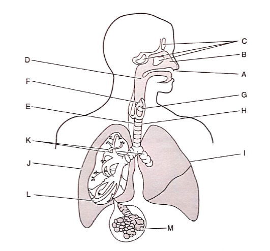
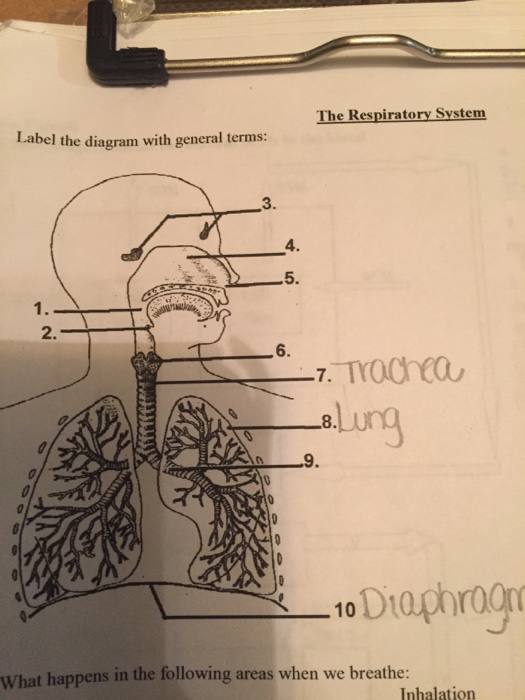
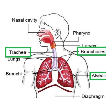
36 label the following diagram of the respiratory system
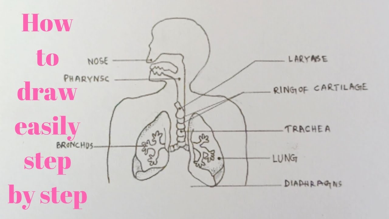
Draw a diagram of human respiratory system and label on it : (a) Diaphragm (b) Larynx. 1 answer. Draw a neat labelled diagram of human respiratory system showing the mechanism of (a) inspiration (b) expiration. asked Feb 10, 2020 in Biology by Santanu01 (51.0k points). The human respiratory system consists of a pair of lungs and a series of air passages leading to the lungs. The entire respiratory tract (passage) Human beings also have a system for transportation of gases. Oxygen is carried by haemoglobin of the red blood cells. Haemoglobin has a great affinity for...
In the throat the trachea or windpipe filters the air. Upper respiratory system science label the diagram as the bronchus enter the lungs t...
Label the following diagram of the respiratory system
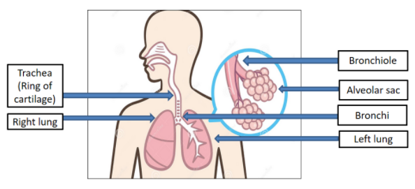
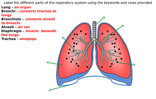
Label your completed diagram. Draw lines away from each structure to an open space using a ruler or straight edge. Clearly label each structure or region correctly. You could also draw a microscope bubble and draw alveoli inside of it to illustrate the smaller structures inside of the lungs. The respiratory system. The following structures can be found in the human thorax Pleural membranes surround each lung. Cartilage rings in the walls of the trachea help to keep it open. The bronchi split into smaller and smaller tubes called bronchioles. Parts of the Respiratory System What is the difference between the two images in terms of the details you canobserve from the subject?b. How does a microscope help in observing and studying things aroundus?c. How will you describe a microscope?
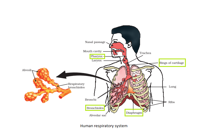
Label the following diagram of the respiratory system. The function of the human respiratory system is to transport air into the lungs and to facilitate the diffusion of oxygen into the blood. Respiratory system diagram. Lower respiratory tract organs. Trachea: Also known as the windpipe this is the tube that carries air from the throat into the lungs. Learn about labeled diagrams respiratory system with free interactive flashcards. Pulmonary ventilation, external respiration, respiratory gas t… the act of bringing air into and out of the lungs and exchangi… Respiratory System Label Diagram Worksheets & Teaching. Education. Details: The respiratory system The human respiratory system medical illustration with internal organs diagram of the respiratory system with labels stock illustrations Lungs with Alveoli Labeled CG image of woman's... Labeled diagram of the lungsrespiratory system. 1024x1024 outline of respiratory system. Human Respiratory System Diagram U...
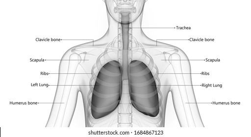
The respiratory system supplies the body with oxygen and disposes of carbon dioxide. There are four processes involved with respiration. These particular squamous cells labeled are going to be called Type 1 alveoli cells of the alveolar wall. The Type 2 cell are green and have a sort of different shape... 1. Add the following labels to the diagram of the respiratory system of a dog below: trachea; bronchioles; diaphragm; bronchi; ribs; larynx; pleural membranes; pleural cavity; rings of cartilage around trachea; alveoli. Label the diagram of the respiratory system below. Lab values for cardiac unite. Respiratory and lung worksheets mrs. Alveoli bronchi bronchioles larynx Similar images Save. lung. The lungs are the primary organs of respiration in humans and many other animals including a few fish and some snails. Diagram of the Human Respiratory System (Infographic). Find out all about your lungs and how breathing works. (Image credit: Ross Toro, Livescience contributor). The primary organs of the respiratory system are the lungs, which function to take in oxygen and expel carbon dioxide as we...
> Draw a diagram of the respiratory system and label the following. (i) Part through which air is taken in. (ii) Part which protects the lungs. (iii) Part which carries the air into the lungs. In the throat the trachea or windpipe filters the air. Once in the lungs oxygen is moved into the bloodstream. How To Draw... Kids will find diagrams of the respiratory system, vocabulary words, review questions Respiratory System: Look at a labeled drawing of the upper and lower respiratory tract by clicking on this link. Respiratory System: Oxygen Delivery System: On this page is a very simple explanation of the... Your respiratory system is the network of organs and tissues that help you breathe. This system helps your body absorb oxygen from the air so The muscles that power your lungs are also part of the respiratory system. These parts work together to move oxygen throughout the body and clean...
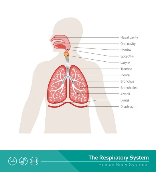
With a labeled diagram, you can see all of the main structures of an organ system together on one page - great for helping you to memorise the appearance of several structures and their relations. Take a look at the labeled diagram of the respiratory system above.
The respiratory system (also respiratory apparatus, ventilatory system) is a biological system consisting of specific organs and structures used for gas exchange in animals and plants.
RESPIRATORY SYSTEM IN ANIMAL( INSECT) Insects breathe using air tubes called trachea. Air vessel out into the trachea through a small hole every body segments called 5. Add the following labels to the diagram below of the urinary system of a mammal. Biology workbook and practical work.
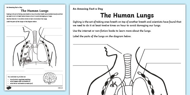
1M followers. In this assignment, students color the various parts of the respiratory system and then answer some follow up questions to describe the functions of the respiratory system.This assignment is geared toward 9th/10th grade Biology students.
Respiratory system quizzes and labeled diagrams. This is a quiz called respiratory system labeling interactive and was created by member teacherrojas advertisement. Draw A Diagram Of Human Respiratory System And Label The Following.
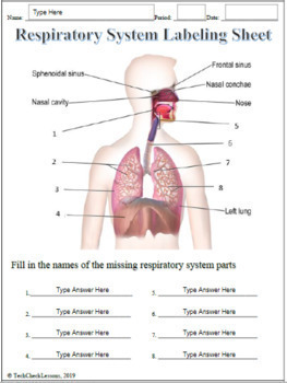
Experts are tested by Chegg as specialists in their subject area. We review their content and use your feedback to keep the quality high. Transcribed image text : Label the diagram of the respiratory system below with the following parts, then colour your diagram.

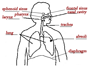
Label the diagram of the respiratory system below. Lab values for cardiac unite. Respiratory and lung worksheets mrs. Alveoli bronchi bronchioles The respiratory tract in humans is made up of the following parts: External nostrils - For the intake of air.; Nasal chamber - which is lined with hair and...
Human respiratory system, the system in humans that takes up oxygen and expels carbon dioxide. The major organs of the respiratory system include the While every effort has been made to follow citation style rules, there may be some discrepancies. Please refer to the appropriate style manual or...

Respiratory System Labeling InteractiveEC. a quiz by teacherrojas. This is an online quiz called Respiratory System Labeling Interactive. There is a printable worksheet available for download here so you can take the quiz with pen and paper.
#respiratorysystemdiagram #humanrespiratorysystemdiagram #respiratorysystem. How to draw human respiratory system/respiratory system/draw labelled diagram of respiratory system. 2 625 просмотров 2,6 тыс. просмотров.
Respiratory System, CFD Simulation and Fluid | ResearchGate, the professional network for scientists. Coandă MAV relates to the physical phenomenon whereby a stream of fluid at high velocity will attach to a curved surface rather than follow its original straight line direction. ...
The respiratory system is an integrated network of organs and tubes that coordinates the exchange of oxygen and carbon dioxide between an organism and its environment. Harmony is seen in the fact that the respiratory system in animals involves the consumption of oxygen and contribution of carbon...

Parts of the Respiratory System What is the difference between the two images in terms of the details you canobserve from the subject?b. How does a microscope help in observing and studying things aroundus?c. How will you describe a microscope?
The respiratory system. The following structures can be found in the human thorax Pleural membranes surround each lung. Cartilage rings in the walls of the trachea help to keep it open. The bronchi split into smaller and smaller tubes called bronchioles.
Label your completed diagram. Draw lines away from each structure to an open space using a ruler or straight edge. Clearly label each structure or region correctly. You could also draw a microscope bubble and draw alveoli inside of it to illustrate the smaller structures inside of the lungs.


0 Response to "36 label the following diagram of the respiratory system"
Post a Comment