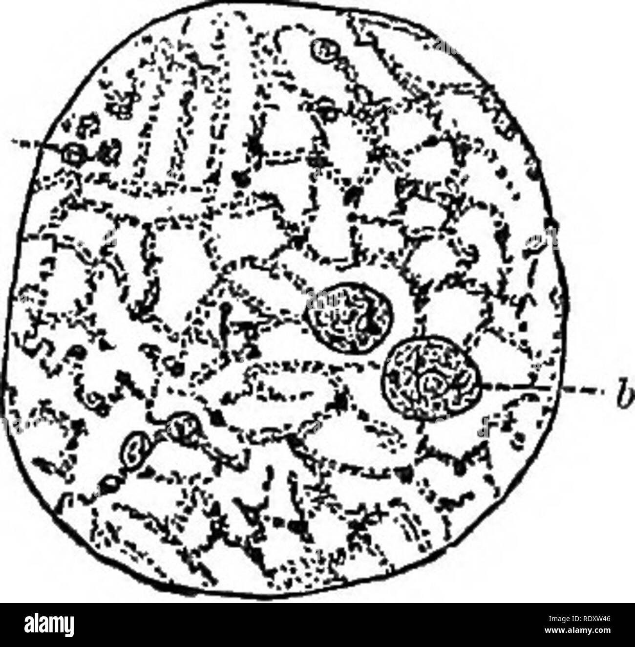
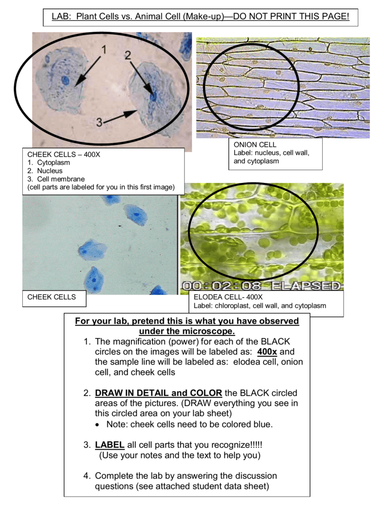
37 diagram of elodea cell
In other words, the primary purpose of plasmodesmata is to develop cell-to-cell communication in plants, similar to gap-junction in animal cells. They are also found in algae. Abundance in Plant Cells. Although they occur in varying numbers, a typical plant cell has between 103 and 105 plasmodesmata, distributed within 1 and 10 per µm 2. Hep2o Hand Titan Plastic Push Fit Straight Tap Connector 15mm X Pipe Fittings Screwfix Com Jual Penyambung Kabel Scotch Terbaru Lazada Co Id Pin On Tools Delta Kitchen Faucet Cartridge Repair Delta Stem Unit Assembly Rp50587 Delta Cartridge Diamond Valve 1h Rep Delta Kitchen Faucet Kitchen Faucet Faucet Repair Washing Machine Diswasher Tee Valve Tap […]
How large is an onion cell? In the diagram to the right, the average size of each cell is 0.25mm. Which is bigger onion cell or elodea cell? An onion cell is approximately 0.13 mm long and . 05 mm wide.

Diagram of elodea cell
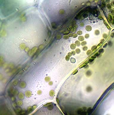
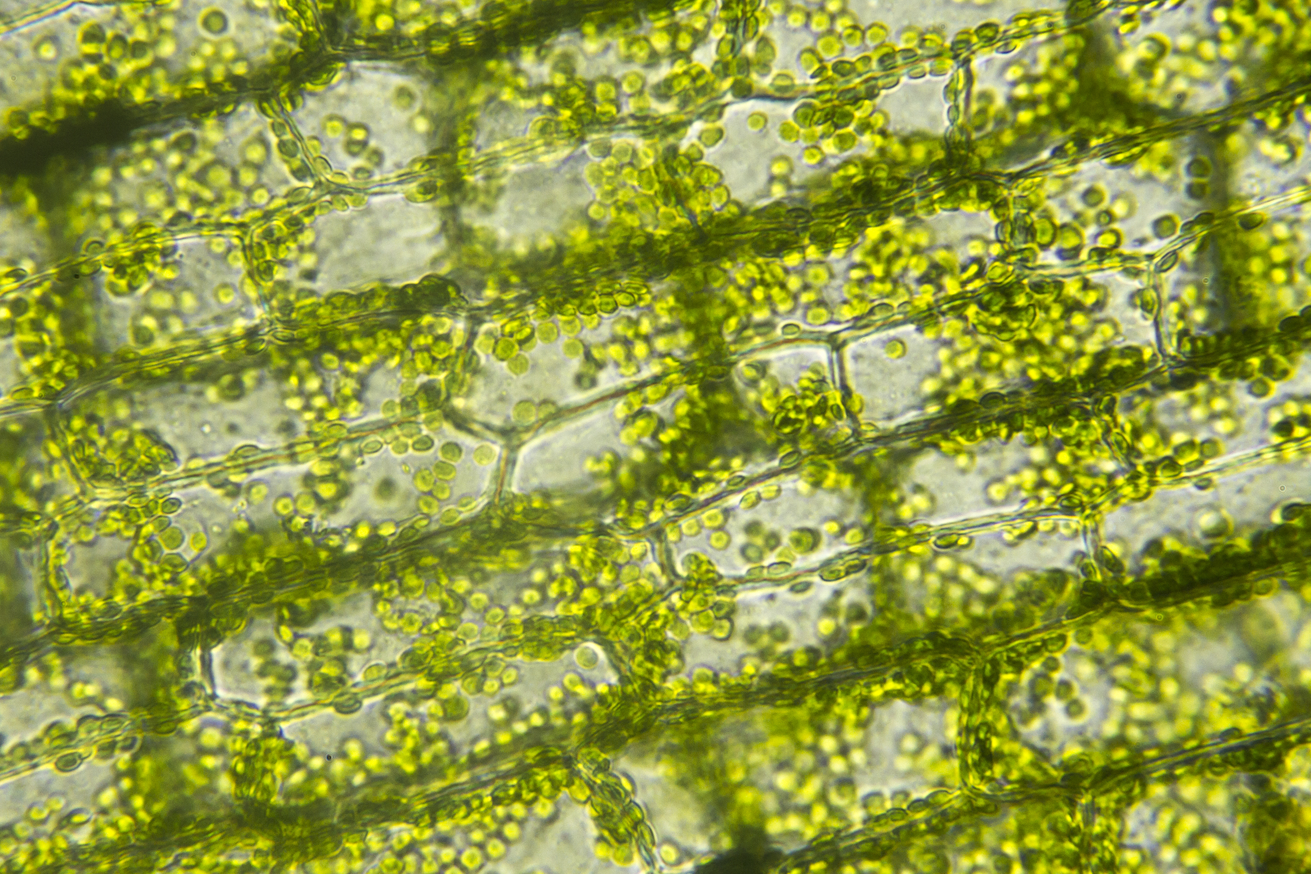
Figure \(\PageIndex{7}\): This image shows cells in the leaf of an aquatic plant, Elodea. Each cell is filled with small green discs which often appear to line the edges of the cell. These are chloroplasts (four are indicated and labeled in the image). Photo credit: Melissa Ha, CC BY-NC. Figure \(\PageIndex{8}\): A diagram of chloroplast anatomy. Students learn a simple technique for quantifying the amount of photosynthesis that occurs in a given period of time, using a common water plant (Elodea). They use this technique to compare the amounts of photosynthesis that occur under conditions of low and high light levels. Before they begin the experiment, however, students must come up with a well-worded hypothesis to be tested. After ... The Elodea leaf consists of two layers of cells. Solely one layer ofcells is in focus when utilizing the excessive energy (40x) goal. Click on to see full reply. Subsequently, one can also ask, how large is an elodea cell? The plasma membrane is just too skinny to see at this magnification. Within the printed picture the scholars work with, the ...
Diagram of elodea cell. The cellular components are called cell organelles. These cell organelles include both membrane and non-membrane bound organelles, present within the cells and are distinct in their structures and functions. They coordinate and function efficiently for the normal functioning of the cell. A few of them function by providing shape and support ... Elodea Leaf Cell Diagram The Elodea leaf is composed of two layers of cells. Only one layer of cells is in focus when using the high. Examining elodea (pondweed) under a compound microscope. solution) and a coverslip and observe the chloroplasts (green structures) and the cell walls. Contractile Vacuole 2 Peace Symbol Biology Symbols. Animal Vs Plant Cells Nice Unlabeled Diagrams Exploring Nature Plant Cell Diagram Cell Diagram Plant Cell. Elodea Water Plant Under Microscope Cell Walls And Chloroplasts Are Clearly Vis Sponsored Microscope Cell Walls E Water Plants Cell Wall Plant Cell. In cell, diagram, elodea. Read Also: Plant cell- definition, labeled diagram, structure, parts, organelles Observation after staining. Under the microscope, plant cells are seen as large rectangular interlocking blocks. The cell wall is distinctly visible around each cell. The cell wall is somewhat thick and is seen rightly when stained.
40x 400x Compound Monocularbiological Microscope45 Degree Angled Headelectric Lightedbeginner Slides Plant Cell Things Under A Microscope Plant Cell Picture. Plasmolysis Of Elodea Biology Experiments Apologia Biology Teaching Science. Elodea Leaf Cell Under Microscope Plant Cell Cells Worksheet Lab Activities. November 16, 2021. Elodea Leaf Cell Under Microscope Plant Cell Cells Worksheet Lab Activities. Cell Transport Lab Osmosis And Diffusion Cell Transport Cell Osmosis. Plant Cells Vs Animal Cells With Diagrams Animal Cell Plant Cell Diagram Plant Cell. Plant Cell Electron Microscope Worksheet Cell Diagram Plant Cell Plant Cell Diagram. Analysis - Venn Diagram 4. The motion is common to ifletype interior of cells and is called cyclonic or cytoplasmic streaming. This appears to be a method of adjusting the amount of damage to the plant by sunlight. Use aesthetic filters to fine tune your search by copy space, frame and duration rates, or depth of filteype. Plant Cell Diagram Under Microscope. For plant cells, there is a cell wall. It's a thin slice: Here's a diagram of a plant cell: The diagram is very clear, and labeled; but at the same time it is interpretive. A bacteria diagram basically facilitates us to profit extra about this unmarried cell organisms that have […]
3. "Animal cells have mitochondria; plant cells have chloroplasts." Is this statement true or false? Explain. 4. Fill out the Venn diagram below to show the differences and similarities between the onion cells and the Elodea cells. Placing Elodea cells into 100% water, which is more hypotonic than freshwater, also causes water movement into of the cells resulting in the swelling of the cells. Thus, a hypertonic solution has less water than the cell and water moves (diffuses) out of the cell by osmosis. Plant Cell Diagram Biology Corner. Images of cells showing the major structures and organelles, including a diagram that maps the process by which proteins are made and exported. Plant Cell Lab - microscope observation of onion and elodea Plant Cell Lab Makeup - can be done at home or at the library Plant Cell Virtual […] Chloroplasts Definition. The word chloroplast is derived from the Greek words chloros, which means green, and plastes, which means "the one who forms".; Chloroplasts are a type of membrane-bound plastids that contain a network of membranes embedded into a liquid matrix and harbor the photosynthetic pigment called chlorophyll.
Within the diagram to the suitable, the typical measurement of every cell is 0.25mm. Moreover, what's the measurement of an onion cell in micrometers? Stained on Excessive Energy (400x), estimated measurement: If subject of view is 0.4 mm (400 µm) and see 2 cells lengthwise: Estimated cell size is 400 µm/2 = 200 µm; width: 400 µm/10 = 40 µm.
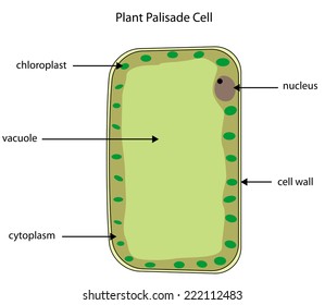
Special Structures in Plant Cells. Most organelles are common to both animal and plant cells. However, plant cells also have features that animal cells do not have: a cell wall, a large central vacuole, and plastids such as chloroplasts.. Plants have very different lifestyles from animals, and these differences are apparent when you examine the structure of the plant cell.
Where is the green color in elodea cells? A group of leaf cells from the pondweed Elodea The green dots are chloroplasts. Notice the colourless space - the vacuole - in the middle of the cells. The nucleus appears to be hidden (between the chloroplasts?). Are all plant cells green in Colour? What are the 10 parts of plant cell?
Chloroplast division occurs every cell cycle right before the separation of two daughter cells (cytokinesis). Cut a small piece of Elodea leaves and put on the slide. How many chloroplasts can be found in one cell? A typical animal cell is 10-20 μm in diameter, which is about one … When thin sections of a chloroplast are examined under the electron microscope, several features are ...
Open Biology Lab Plasmolysis Osmosis In Elodea And Potato Cells Biology Labs Cell Transport Biology Classroom Onion And Cheek Cell Lab Biology Labs Lab Report Lab Report Template Stellar Classification and the H-R Diagram 1 Introduction Stellar Classi cation As early as the beginning of the 19th century scientists have studied absorption ...
Plasmolysis In Plant Cell Diagram. The cells with only cell membranes, such as animal cells, eventually swell and burst. This indicates the phenomenon of deplasmolysis. A bacteria diagram clearly enables us to profit extra about this unmarried cell organisms that have neither membrane-bounded nucleolus or organelles like mitochondria and chloroplasts. They are obviously a trigger […]
Mar 26 2020 Elodea is a water plant. Histopathology lab is the place where the specimen gets processed and stained to view under Procedure To view the yogurt culture the yogurt has to sit in a dark warm place for 12 24 hours before it is viewed under a microscope. ... After that they started to circulate through the cell body. The diagram shows ...
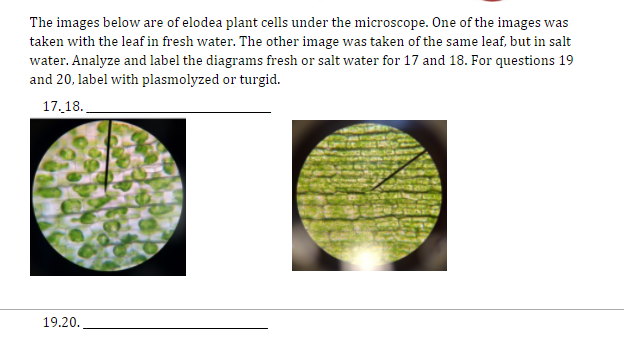
Water will flow out of the Elodea cells by osmosis, shrinking the cell membrane away from the stiff cell wall (plasmolysis). Get a microscope slide. Place 2 drops of dI water on the left and 2 drops 20% salt on the right. Obtain a leaf from a stalk of Elodea and cut the leaf in half. Place a half leaf in each solution.
Elodea Cell Diagram. abby.kulas July 20, 2021 Templates No Comments. 21 posts related to Elodea Cell Diagram. Froth Flotation Cell Diagram. Att Cell Tower Map Ny. Map Of Cell Coverage. Att Cell Tower Map Georgia. Att Cell Tower Mapper. Cell Phone Coverage Map Nz. Cell Phone Tracking Map.
The Elodea leaf consists of two layers of cells. Solely one layer ofcells is in focus when utilizing the excessive energy (40x) goal. Click on to see full reply. Subsequently, one can also ask, how large is an elodea cell? The plasma membrane is just too skinny to see at this magnification. Within the printed picture the scholars work with, the ...
Students learn a simple technique for quantifying the amount of photosynthesis that occurs in a given period of time, using a common water plant (Elodea). They use this technique to compare the amounts of photosynthesis that occur under conditions of low and high light levels. Before they begin the experiment, however, students must come up with a well-worded hypothesis to be tested. After ...
Figure \(\PageIndex{7}\): This image shows cells in the leaf of an aquatic plant, Elodea. Each cell is filled with small green discs which often appear to line the edges of the cell. These are chloroplasts (four are indicated and labeled in the image). Photo credit: Melissa Ha, CC BY-NC. Figure \(\PageIndex{8}\): A diagram of chloroplast anatomy.






/plant-cell-elodea--isotonic-solution-shows-cells--chloroplasts-250x-at-35mm-139802547-5a956de86bf069003717851a.jpg)








0 Response to "37 diagram of elodea cell"
Post a Comment