35 drag the labels onto the diagram to identify the various muscle structures.
Drag the labels onto the diagram to identify the various chromosome structures. Drag the labels to the correct locations on these images of human chromosomes. Drag the labels onto the diagram to identify the stages of the life cycle. (Drag only blue labels onto blue targets and pink labels onto pink targets.) The function(s) of meiosis is/are ... Drag the labels onto the flowchart to identify the steps of the sliding filament model of muscle contraction. 1 the myosin head binds the actin. Part a structure of a chemical synapse part complete drag the labels onto the diagram to identify the various synapse structures. How synapses work synapse structure 6 of 12 complete can you identify ...
Principles and Techiniques of Biochemistry and Molecular Biology 7th ed wilson walker

Drag the labels onto the diagram to identify the various muscle structures.
Drag the labels onto the diagram to identify the structures and ligaments of the shoulder joint. (musculotendinous cuff), the circle of tendons around the shoulder joint. Acomioclavicular lgament glenohumeral ilgaments glenoid cavily glenoid iabrum tendon of biceps brachii muscle articular capsule coracohumera ligament. 9 Jul 2021 — The coracohumeral, glenohumeral ligaments and the tendons of the supraspinatus and subscapularis muscles all serve to support and strengthen. If ... 21 Apr 2021 — The coracohumeral, glenohumeral ligaments and the tendons of the supraspinatus and subscapularis muscles all serve to support and strengthen.
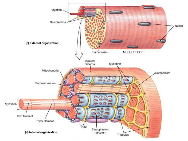
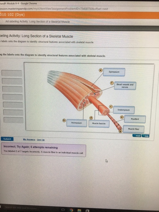
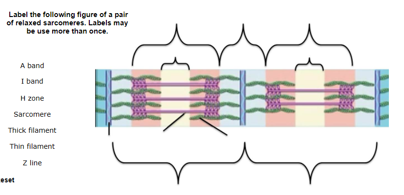
Drag the labels onto the diagram to identify the various muscle structures.. Drag the labels onto the flowchart to identify the sequence of steps that occurs during muscle contraction. The step numbers correspond to the numbers in the diagram above. ANSWER: Reset Help Myosin heads bind to actin and shorten the sarcomere. Action potential spreads down tubules in the muscle cell. Drag the labels onto the diagram to identify the various muscle structures. a) Muscle b) Bundle of muscle fibers c) Single muscle fiber (cell) d) Myofibril View Muscle_Structure_Label.png from BIOLOGY 107 at Embry-Riddle Aeronautical University. Part A - Structure Of a ITIUSCE Drag the labels onto the diagram to identify the various muscle The structure of a skeletal muscle fiber drag the labels onto the diagram to identify structural features associated with a skeletal muscle fiber. These tissues include the skeletal muscle fibers blood vessels nerve fibers and connective tissue.
In chapter 3, we learned that the various compartments have differing ... Drag the labels onto the diagram to identify parts of the neuromuscular junction. Rating: 5 · 3 reviews BioFlix Activity: Muscle Contraction -- Muscle Structure Part A - Structure of a muscle Drag the labels onto the diagram to identify the various muscle structures. Reset Help Sarcomere Myofibril Muscle Single muscle fiber (cell) Myosin (thick filaments) Actin (thin filaments) AASTA Bundle of muscle fibers Submit Request Answer. Drag the labels onto the diagram to identify the various types of cells in the retina. In a condition called detached retina, the neural layer of the retina separates from the pigmented part. Blindness may result if blood supply to the photoreceptors cannot be restored. BioFlix Activity: Muscle Contraction -- Muscle Structure Part A - Structure of a muscle Drag the labels onto the diagram to identify the various muscle structures. Reset Help Sarcomere Myofibril Muscle Single muscle fiber (cell) Myosin (thick...
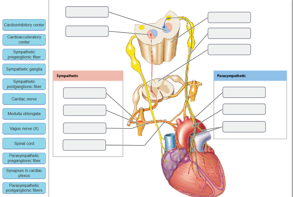
Drag the labels on the left onto the diagram of the animal cell to correctly identify the function performed by each cellular structure. a. synthesizes lipids b. assembles ribosomes c. defines cell shape d. produces secretory proteins e. modifies and sorts proteins f. digests proteins g. generates ATP The eukaryotic cell has well-defined structures that serve discrete functional … This problem has been solved. Drag the labels onto the diagram to identify the parts of the cell. Drag the labels onto the diagram to identify the mechanisms involved in the transport of carbon d. Part a animal cell structure drag the labels onto the diagram to identify the structures of an ani. Drag the labels onto the diagram to identify the muscle types based on fascicle organization. look at pic Drag the labels onto the diagram to identify the major skeletal muscles, anterior view. Drag the labels onto the diagram to identify the components of the autonomic nervous system. visceral effectors: ( top down) 1. smooth muscle 2. glands. 3. cardiac muscle 4. Adipocytes top middle to bottom middle 1. visceral motor nuclei in hypothalamus 2. autonomic ganglia 3. ganglionic neurons 4. preganglionic neuron
25 Drag The Labels Onto The Diagram To Identify Parts Of ...
Identify structures of a generic neuron on a model or diagram ... Each skeletal muscle is an organ that consists of various integrated tissues.

Drag The Labels Onto The Diagram To Identify Structural ...
Drag The Labels Onto The Diagram To Identify The from lh5.googleusercontent.com Drag the labels onto the diagram to identify the various types of synarthroses and. Joint capsule * strong * reinforced by capsular ligaments * only place where shoulder girdle attaches to axial . Drag the labels onto the diagram to identify the structures and ...

27 Drag The Labels Onto The Diagram To Identify Structural ...
Academia.edu is a platform for academics to share research papers.

Drag The Labels Onto The Diagram To Identify The ...
Drag the labels onto the diagram to label the steps of smooth muscle activation and deactivation. Q. Skeletal muscle is capable of which of the following?a. wave summationb. fused tetanusc. autorhythmicityd. wave summation and fused tetanuse. wave sum... Q. Explain why, during exercise, muscle cells need liver cells for regeneration of glucose ...
Solved: Drag The Labels Onto The Diagram To Identify The C ...
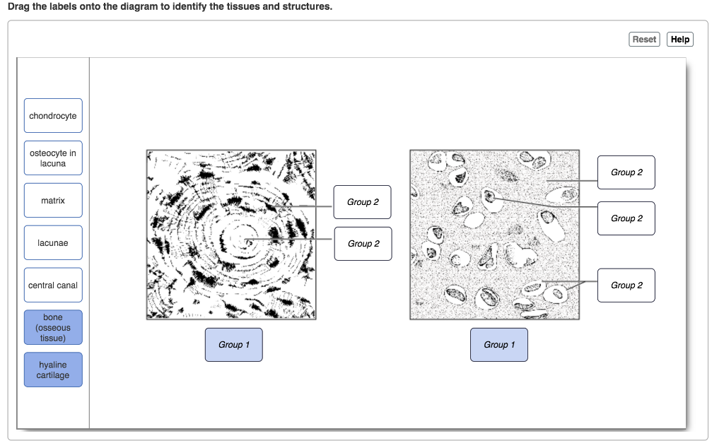
Drag the labels onto the diagram to identify the various chromosome structures. Start studying cell cycle labeling. Prophase chromatids nucleolus telophase chromosome chromatin interphase spindle fibers centrioles metaphase nuclear membrane daughter cells anaphase cell membrane centromere use your notes and textbook to answer the questions below.

Drag The Labels Onto The Diagram Of Muscle Spindle ...
Drag the labels onto the diagram to identify the various chromosome structures. Then drag white labels onto white targets only to identify the ploidy level at each stage. Help reset cytokinesis mitosis phase mitotic phase phase interphase phase. Drag the labels onto the diagram to identify aspects of gas transport and exchange.

Fit man
BioFlix Activity: Muscle Contraction -- Muscle Structure Part A - Structure of a muscle Drag the labels onto the diagram to identify the various muscle structures. Reset Help Sarcomere Myofibril Muscle Single muscle fiber (cell) Myosin (thick filaments) Actin (thin filaments) AASTA Bundle of muscle fibers Submit Request Answer.
Solved: Drag The Labels Onto The Diagram To Identify The T ...
Anatomy and physiology 2Drag the labels onto the diagram to identify the parts of the pituitary gland & its associated structures
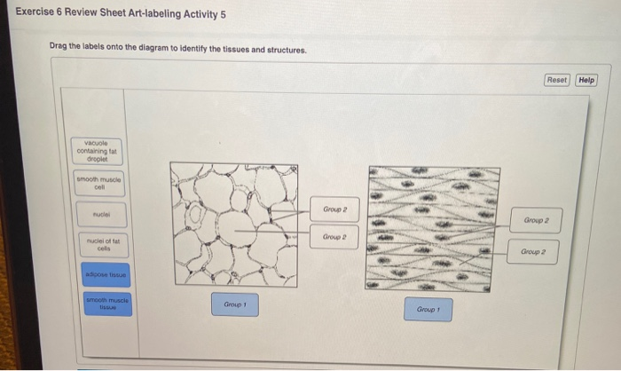
Solved: Exercise 6 Review Sheet Art-labeling Activity 5 Dr ...
21 Apr 2021 — The coracohumeral, glenohumeral ligaments and the tendons of the supraspinatus and subscapularis muscles all serve to support and strengthen.
Drag The Labels Onto The Diagram To Identify Parts Of The ...
9 Jul 2021 — The coracohumeral, glenohumeral ligaments and the tendons of the supraspinatus and subscapularis muscles all serve to support and strengthen. If ...
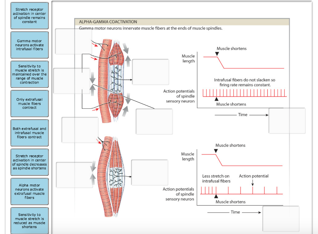
Solved: Label The Structures Involved In Muscle Spindle Fu ...
Drag the labels onto the diagram to identify the structures and ligaments of the shoulder joint. (musculotendinous cuff), the circle of tendons around the shoulder joint. Acomioclavicular lgament glenohumeral ilgaments glenoid cavily glenoid iabrum tendon of biceps brachii muscle articular capsule coracohumera ligament.

30 Drag The Labels Onto The Diagram To Identify Structural ...

29 Drag The Labels Onto The Diagram To Identify The ...
30 Drag The Labels Onto The Diagram To Identify Structural ...
31 Drag The Labels Onto The Diagram To Identify The ...

Cutting Through
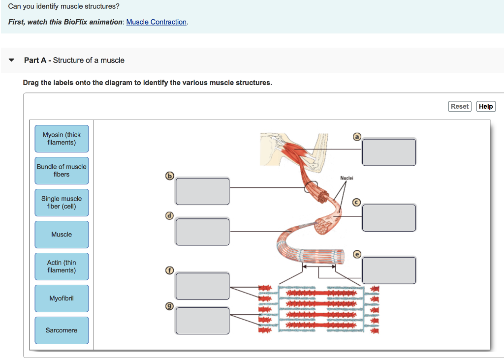
Solved: Can You Identify Muscle Structures? First, Watch T ...
Drag The Labels Onto The Diagram To Identify Structural ...

Chapter 16 Neural Integration II: The Autonomic Nervous ...
Solved: Drag The Labels Onto The Diagram To Identify The T ...
Drag The Labels Onto The Diagram To Identify Structural ...
Drag The Labels Onto The Diagram To Identify The Various ...
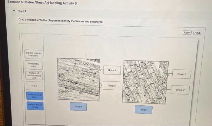
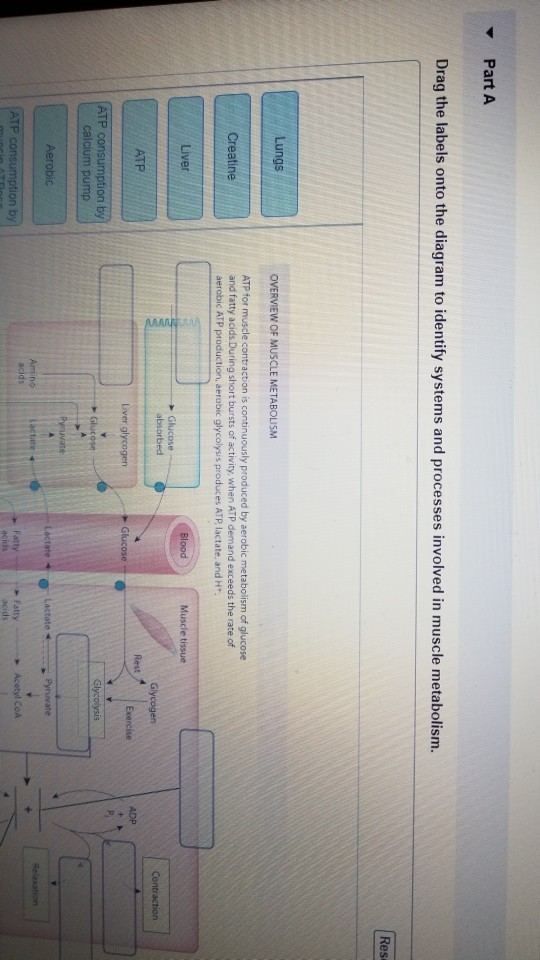
Solved: Exercise 6 Review Sheet Art-labeling Activity 6 Pa ...
Solved: Part A Drag The Labels Onto The Diagram To Ident ...
32 Drag The Labels Onto The Diagram Of Muscle Spindle ...
Drag The Labels Onto The Diagram To Identify The Various ...

Drag The Labels Onto The Diagram To Identify The Various ...
32 Drag The Labels Onto The Diagram To Identify Structural ...

Desfile del Orgullo Gay de Torremolinos 2019

Drag The Labels Onto The Diagram To Identify Structural ...
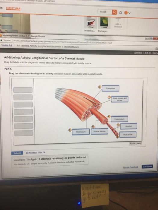
Solved: -Rea-muscle-q15595905 Search Add A Sextook Modifie ...

Senior bodybuilding right side chest

30 Drag The Labels Onto The Diagram To Identify Structural ...

Drag The Labels Onto The Diagram Of Muscle Spindle ...

Drag The Labels Onto The Diagram To Identify The ...
0 Response to "35 drag the labels onto the diagram to identify the various muscle structures."
Post a Comment