39 nerves in the head diagram
There are four pairs of sinuses (named for the skull bones in which they're located). Interactive diagrams show sinus cavity locations and help visualize sinusitis, the most common type of sinus in... The nerves of the cervical plexus supply the back of the head, the neck and the shoulders. Brachial Plexus The arrangement of nerve fibers formed by the ventral rami of the lower cervical and upper thoracic nerve root, precisely between the nerve roots of the 5th cervical and 1st thoracic vertebra, is known as the brachial plexus.
Anatomical Course The vagus nerve has the longest course of all the cranial nerves, extending from the head to the abdomen. Its name is derived from the Latin... In the Head The vagus nerve originates from the medulla of the brainstem. It exits the cranium via the jugular foramen, with the glossopharyngeal and accessory nerves... Log In Subjects Quizzes Pricing Our Apps Contact Us... The Head The Cranial Nerves The Vagus Nerve (CN X)...

Nerves in the head diagram
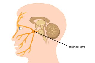
The sensory cranial nerves are involved with the senses, search as sight, smell, hearing, and touch. Whereas the motor nerves are responsible for controlling the movements and functions of muscles and glands, cranial nerves supply sensory and motor information to areas of the head and neck. One nerve, the vagus nerve, extends beyond the neck to ... The trigeminal nerve is the largest and most complex of the 12 cranial nerves (CNs). It supplies sensations to the face, mucous membranes, and other structures of the head. Nerves within the Cervical Spine: Neck Anatomy Nerves Picture. There are 8 spinal nerves that originate from the cervical spine. The majority of these nerves control the functions of the upper extremities and allow you to feel your arms, shoulder, and back of your head. Each nerve provides sensation to a specific area of the body called a ...
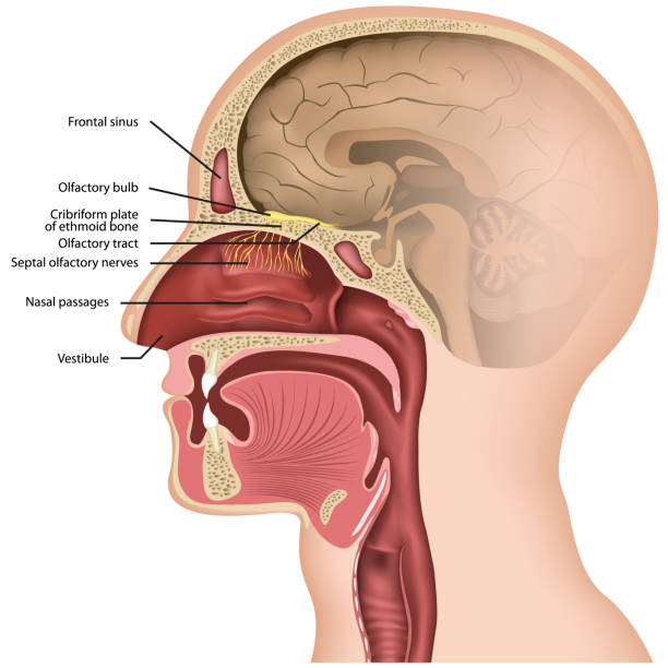
Nerves in the head diagram. Cranial Nerve Anatomy / Cranial nerves. Cell bodies located in the olfactory mucosa overlying the superior nasal concha and superior septum. Axons pass through the cribiform plate of the ethmoid bone. Forms olfactory bulb that connects to the brain via the olfactory tract. Dec 19, 2017 · Because the brain processes all of the body’s signals, it houses major nerves to collect the information and get it to the proper section of the brain. There are 12 pairs of major nerves called ... Diagram Of The Cutaneous Nerves Of The Head And Neck. (Photo By Encyclopaedia Britannica/UIG Via Getty Images) Diagram Of The Cutaneous Nerves Of The Head And Neck. : News Photo. You have view only access under this Premium Access agreement. Contact your company to license this image. Occipital Neuralgia is a condition in which the occipital nerves, the nerves that run through the scalp, are injured or inflamed.This causes headaches that feel like severe piercing, throbbing or shock-like pain in the upper neck, back of the head or behind the ears.
Two spinal nerves branch off from the right and left sides of the spinal cord or the cauda equina at each spinal segment. These spinal nerves are formed by 2 types of fibers—sensory fibers that send messages to the brain (feeling pain when the leg is hurt) and motor fibers that receive messages from the brain (lifting the leg to get out of a car). The nerves of the head include the sympathetic and parasympathetic innervation to the head and neck, as well as the three branches of the trigeminal nerve: ophthalmic, maxillary and mandibular.. The sympathetic innervation begins in the spinal cord.Nerve fibres exit the spinal cord and enter the sympathetic chain, which is composed of superior, middle and inferior cervical ganglion. Sep 13, 2021 · This human anatomy module is about the cranial nerves. It consists of 15 vector anatomical drawings with 280 anatomical structures labeled. It is intended for the use of medical students working on human anatomy, student nurses, physiotherapists, electro-radiological technicians and residents – especially those working in neurology, neurosurgery, otolaryngology – and for any physician ... Instant anatomy is a specialised web site for you to learn all about human anatomy of the body with diagrams, podcasts and revision questions
C1, C2, and C3 (the first three cervical nerves) help control the head and neck, including movements forward, backward, and to the sides. 1 The C2 dermatome handles sensation for the upper part of the head, and the C3 dermatome covers the side of the face and back of the head. 2 (C1 does not have a dermatome.) See The C1-C2 Vertebrae and Spinal ... Possible Implications of Faulty US Technical Intelligence in the Damascus Nerve Agent Attack of August 21, 2013 Richard Lloyd Former UN Weapons Inspector Tesla Laboratories Inc.|Arlington, VA Voice: 509-979-3995; e-mail: ******@*****.*** Theodore A. Postol Professor of Science, Technology, and National Security Policy... Cervical nerves are spinal nerves that arise from the cervical region of the spinal cord. These nerves conduct motor and sensory information via efferent and afferent fibers, respectively, to and from the central nervous system. While classified as peripheral nerves, the motor cell body resides in the anterior horn of the spinal cord. There are eight pairs of cervical nerves, denoted C1 to C8 ... In simple terms, the body is telling the brain that there is a threat and it should alert itself. Vagus nerve vector illustration. Labelled anatomical structure scheme and location diagram of human body longest nerve. Infographic with isolated ganglion, branches and plexus. Inner biological ANS. Source: Shutterstock The nervous...
cutaneous nerve | physiology | Britannica
You probably could implant something in the head [of the size shown in the film]…” Vaisman trails off, stuck on the idea that there’s not much chance of it also containing a battery. “Right now, with things like nerve stimulators that you can put in after a stroke or for Parkinson’s, the battery pack they insert...

Brain Models
The peptide would enable surgeons to see even the most sensitive nerves instead of relying on their experience and electronic monitoring.

Back Of The Head Muscle Structure And Nerve System Diagram ...
The frontal lobe, which is located in the front of the head, is the largest section of the brain. It plays a role in many conscious functions, including personality and movement. It also helps the brain interpret smells. Positioned near the back of the brain, the occipital lobe primarily interprets vision signals. Located on...
File:Head facial nerve branches.jpg - Wikimedia Commons
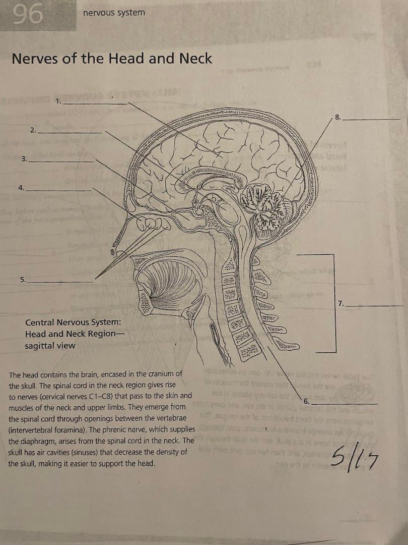
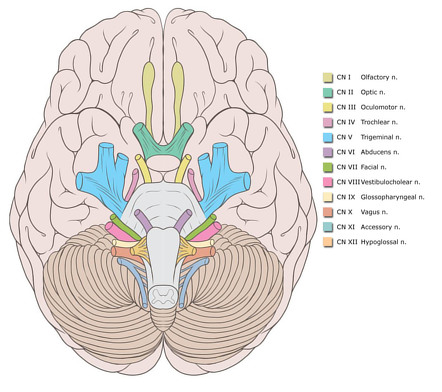
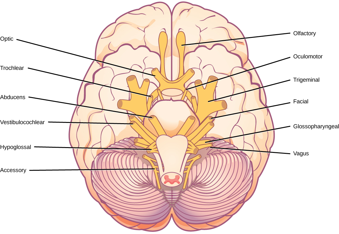
Twelve pairs of cranial nerves arise in the brain and give off branches to the structures of the head and face. These nerves leave the cranial cavity through foramina in the base of the cranium. The fifth cranial nerve (the trigeminal nerve) is the largest of the twelve pairs. See figure 2-13.
/GettyImages-1092334754-fd0644493b3148288970e38fd26aead0.jpg)
Brainstem: Anatomy, Function, and Treatment
Nov 02, 2020 · The nerves of the head and neck include the most vital and important organs of the nervous system — the brain and spinal cord — as well as the organs of the special senses. In addition, in this region we also find the major cranial and spinal nerves that connect the central nervous system to the organs, skin, and muscles of the head and neck.
Trigeminal neuralgia - NHS
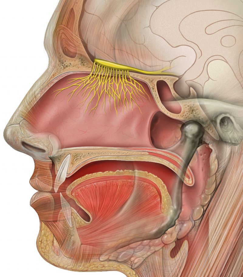
The olfactory nerve is responsible for the sense of smell. The nasociliary and nasopalatine nerves provide general sensation. Anatomical Course The anatomical... common cold) Permanent anosmia can be caused by head injury, or tumours which occur in the olfactory groove (e.g. meningioma). Anosmia can also occur as a result of... Log In Subjects Quizzes Pricing Our Apps Contact Us... The Head The Cranial Nerves The Olfactory Nerve (CN I)...

The brain and nerves of the head and neck - Stock Image ...
Like the lumbar spinal nerves, you also have five pairs of sacral spinal nerves. They’re associated with your , which is one of the bones found in your pelvis.. You only have a single pair of coccygeal spinal nerves. This pair of nerves originates from the area of your, or tailbone.Each of your dermatomes is associated with a...

The 12 Cranial Nerves and their Functions | Medical Library
The Cranial Nervous System nerves connect the brain to the eyes, mouth, ears and other parts of the head. The Autonomic Nervous System nerves connect the central nervous system to the lungs, heart ...

12 Cranial Nerves: Functions & Diagram of Locations | Simply ...
Each nerve also has a descriptive name (e.g. olfactory, optic, etc.) that identifies its function or location. The cranial nerves provide a direct connection to the brain for the special sense organs, muscles of the head, neck, and shoulders, the heart, and the GI tract. Spinal Nerves. Extending from the left and right sides of the spinal cord ...

Cranial nerves of the brain. The cranial nerves provide motor ...
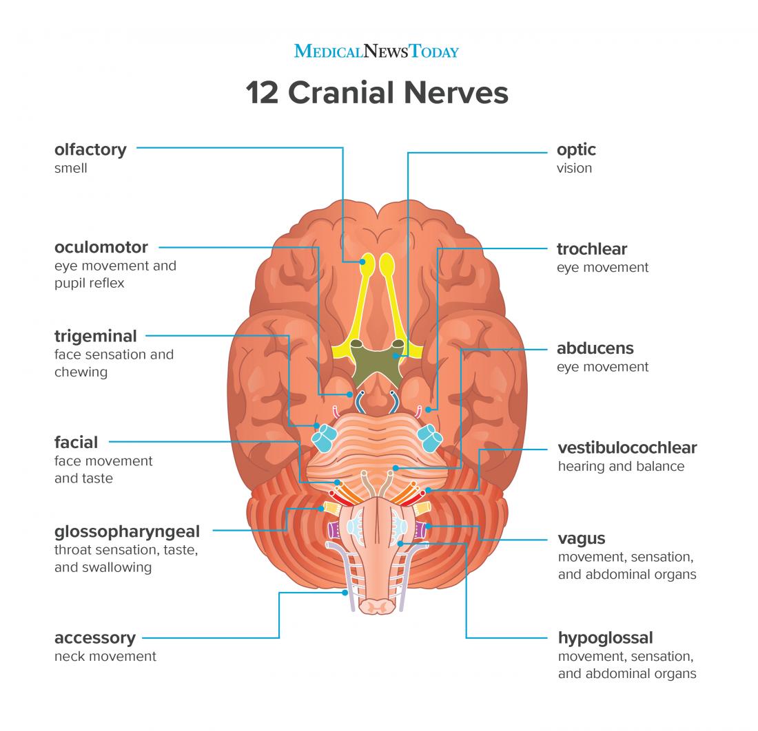
These Are the 12 Cranial Nerves and Their Functions The 12 Cranial Nerves I. Olfactory nerve II. Optic nerve III. Oculomotor nerve IV. Trochlear nerve V. Trigeminal nerve VI. Abducens nerve VII....
Solved 96 nervous system Nerves of the Head and Neck 8. 2 ...
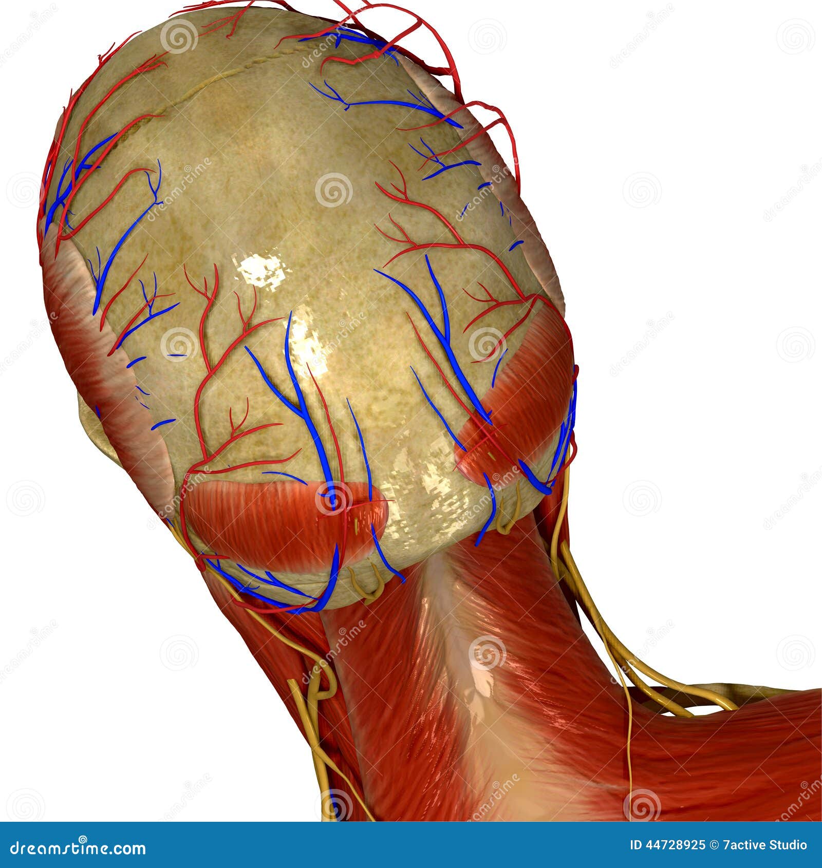
In red are the deep muscles that control your head on your neck. In yellow, you see the following nerves: GON – Greater occipital nerve – This along with the TON originate from the C2 spinal nerve. The GON comes exits alongside the upper trapezius (not shown as that muscle is more superficial than the deep ones illustrated above).
Facial Nerves Stock Illustrations – 103 Facial Nerves Stock ...
Chordate evolution and the origin of craniates: An old brain in a new head

Sensory innervation of the scalp. Lateral view of the head ...
Nicole Long Date: January 29, 2022 A diagram showing nerves in the head and neck.. Nerves in the neck, medically referred to as the cervical spine, help transmit information along the pathways of the central and peripheral nervous system, including sensory and motor skills processes.The cervical spine consists of eight different sets of nerves.

Fig. 449. Nerves of the head. A treatise on human ...
outline the evaluation process and differential diagnosis of nerve injuries to the upper extremity.Axillary Nerve: Quadrilateral Space Syndrome ...

Cranial nerves - Wikipedia
ADVERTISEMENTS: The regulation of the heart is effected through the afferent (centripetal) and efferent (centrifugal) nerves of the heart (Fig. 7.79). The afferent nerves are: ADVERTISEMENTS: i. From the heart through the vagus nerve and from the aortic arch, the aortic nerve. ii. From the heart through the inferior cervical and first four thoracic ganglia […]

Head, Face, and Neck Nerves, labeling and function Diagram ...
12 cranial nerves (diagram) Cranial nerves are peripheral nerves that emerge from the cranial nerve nuclei of the brainstem and spinal cord. They innervate the head and neck. Cranial nerves are numbered one to twelve according to their order of exit through the skull fissures.

Cutaneous innervation head and neck - UpToDate
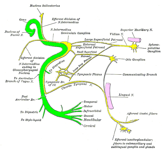
It communicates sensory information from the: outer part of the ear lower part of the mouth and the associated mucous membranes front and middle parts of the tongue teeth of the lower jaw and the associated mucous membranes It also stimulates movement of the muscles in the jaw and some of the muscles within the inner ear.
Cranial nerves | Radiology Reference Article | Radiopaedia.org
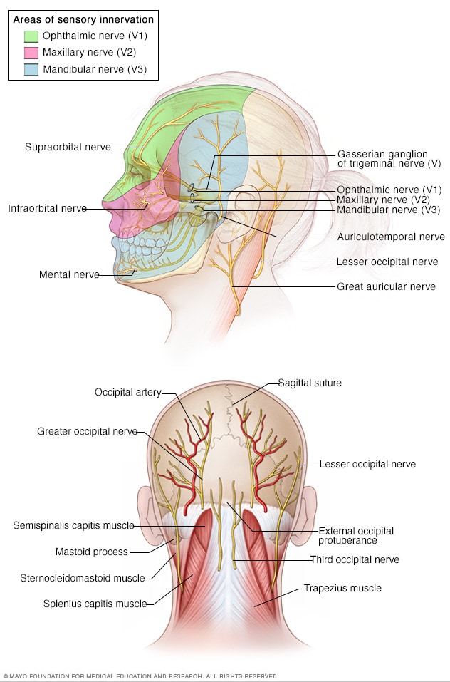
The occipital nerves are a group of nerves that arise from the C2 and C3 spinal nerves.[1][2] They innervate the posterior scalp up as far as the vertex and other structures as well, such as the ear.[2] There are three major occipital nerves in the human body: the greater occipital nerve (GON), the lesser (or small) occipital nerve (LON), and the third (or least) occipital nerve (TON).
The nerves of the head and neck | Anatomy of the nerves of ...
The functions of the cranial nerves are sensory, motor, or both: Sensory cranial nerves help a person to see, smell, and hear. Motor cranial nerves help control muscle movements in the head and neck.

Dog head nerves Diagram | Quizlet
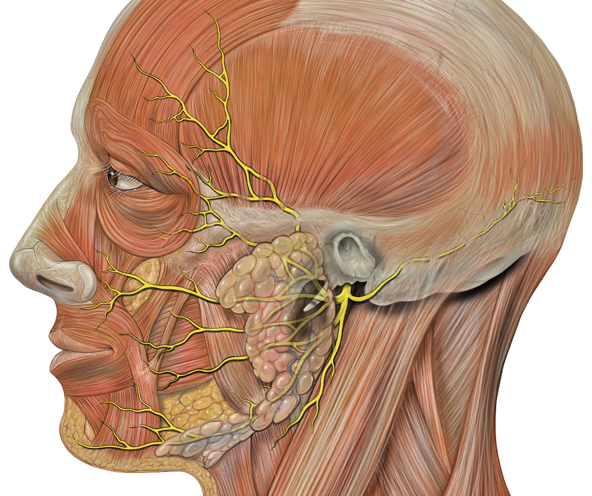
The facial nerve and its branches regulate a number of functions of the mouth and face. Most of its divisions stimulate muscles that allow eyelids to open and close, as well as facial movements. This nerve also mediates the production of tears and saliva and perception of taste in the tongue and receives some sensory input from the face as well ...
Trigeminal neuralgia - Symptoms and causes - Mayo Clinic
The nervous system consists of the brain, spinal cord, and nerves. This is the way the body communicates with the brain and vice versa. The nervous system is divided into two key parts: C entral ...
Your Complete Guide to Trigeminal Neuralgia; A. M. Kaufmann ...
MCQs Audio Podcasts Lectures iPad/iPhone apps About Subscribe Video podcasts Questions Android apps HEAD & NECK THORAX ABDOMEN UPPER LIMB LOWER LIMB Body Head and Neck Nerves Autonomic Cervical ganglia in neck Home Sitemap Privacy Policy - Contact Us Copyright © 1999-2020 - Instant Anatomy
16.4 The Peripheral Nervous System – Concepts of Biology ...
A nerve is a cable-like structure within the body designed to conduct nerve impulses that relay information from one part of the body to another. A typical nerve is made up of a bundle of fibres which are wrapped around layers of tissue and fat, and they stretch throughout the body. These nerves transmit information along the axons to the ...

Nerve Blocks of the Face - NYSORA
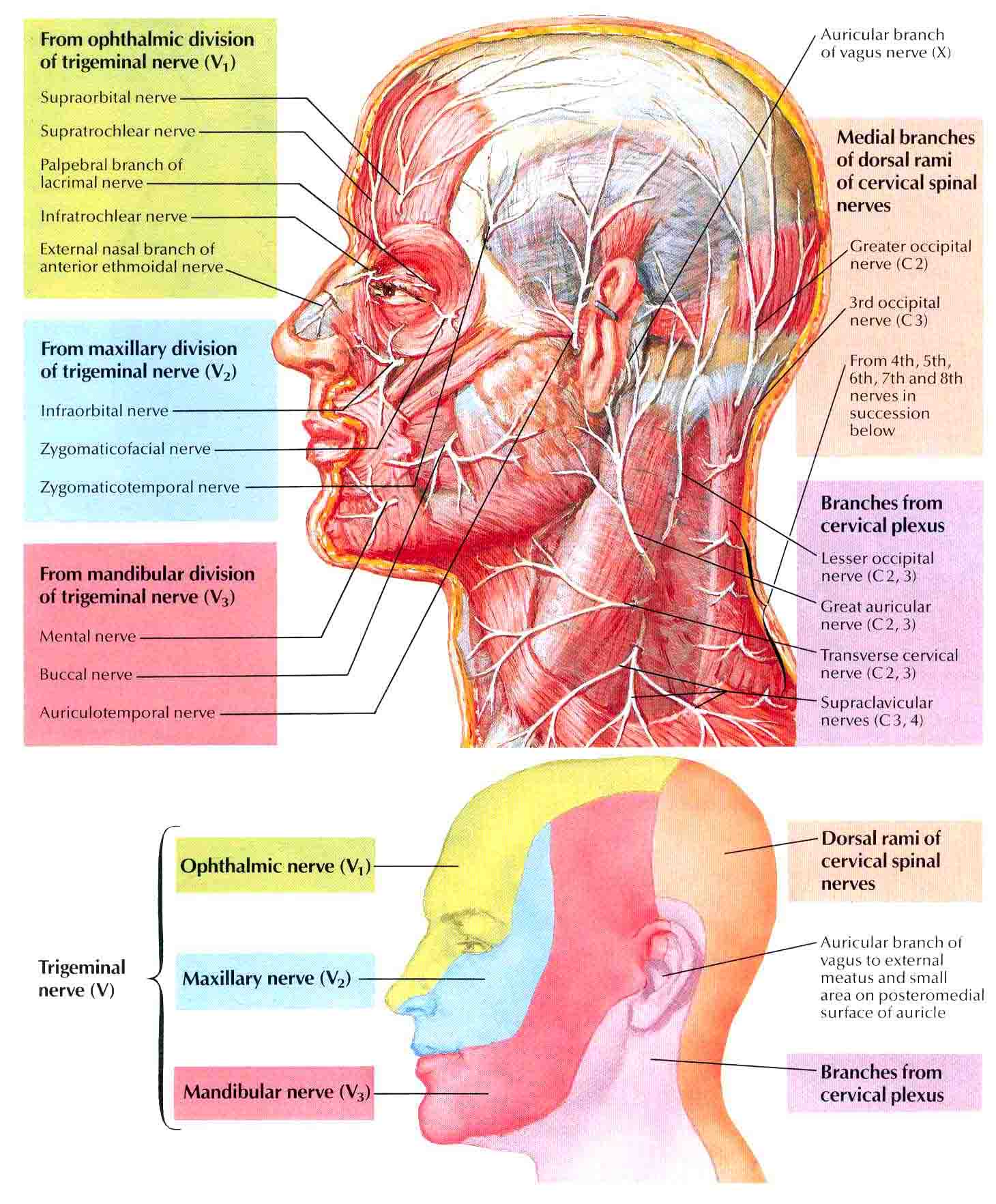
The superficial nerves of the face and scalp are derived from three sources located in the head and neck:. Facial nerve (CN VII), which provides motor innervation to the muscles of the face; Trigeminal nerve (CN V), which provides sensory innervation to the face via its ophthalmic division (CN V1), maxillary division (CN V2) and mandibular division (CN V3)
Cranial Nerve Anatomy / Cranial nerves | Iowa Head and Neck ...
Oct 28, 2021 · The facial nerve provides motor innervation to the muscles of facial expression. Salivary glands are controlled by autonomic nerves stemming mainly from the same facial nerve. The cervical plexus is formed by the C1 to C5 spinal nerves, giving off two branches innervating the head: lesser occipital and greater auricular nerves.
Nerves and arteries of head and neck: Anatomy, branches | Kenhub
Peripheral (outside of the central nervous system) nerves are tubes that are special in their ability to transmit electric impulses along their length and into or away from the central nervous system. Nerves have specialized receptors for different inputs like hot, cold, sharp and vibration. Smaller nerves are grouped into larger rope-like groups that travel up and down the body.

Diagram of the cutaneous nerves of the head and neck. Large ...
Nerves within the Cervical Spine: Neck Anatomy Nerves Picture. There are 8 spinal nerves that originate from the cervical spine. The majority of these nerves control the functions of the upper extremities and allow you to feel your arms, shoulder, and back of your head. Each nerve provides sensation to a specific area of the body called a ...
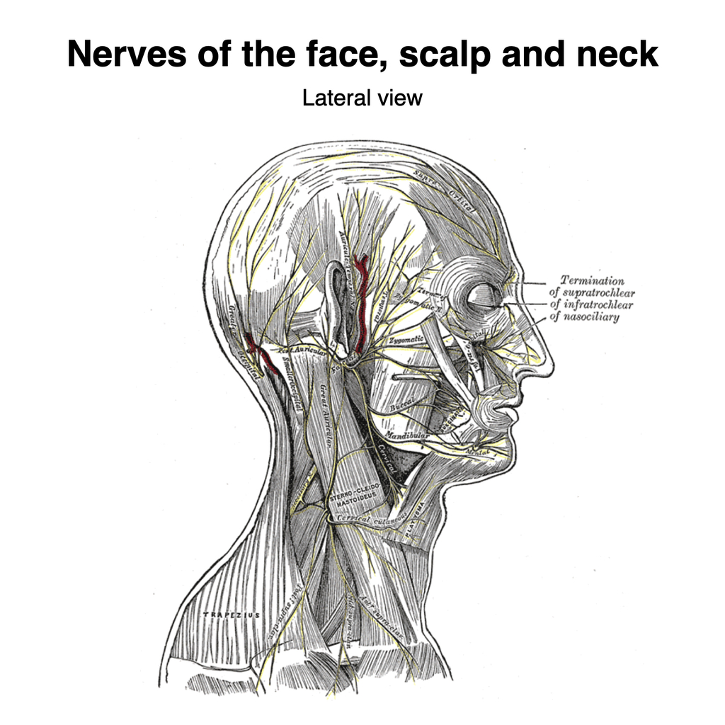
Nerves of the face, scalp and neck (Gray's illustration ...
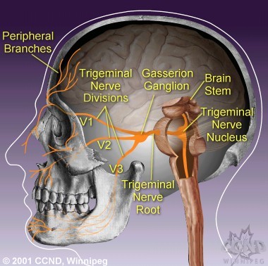
The trigeminal nerve is the largest and most complex of the 12 cranial nerves (CNs). It supplies sensations to the face, mucous membranes, and other structures of the head.
15.2: Cranial Nerves - Biology LibreTexts
The sensory cranial nerves are involved with the senses, search as sight, smell, hearing, and touch. Whereas the motor nerves are responsible for controlling the movements and functions of muscles and glands, cranial nerves supply sensory and motor information to areas of the head and neck. One nerve, the vagus nerve, extends beyond the neck to ...

Nerves and vessels of the head, illustration - Stock Image ...
What are the 12 cranial nerves? Functions and diagram
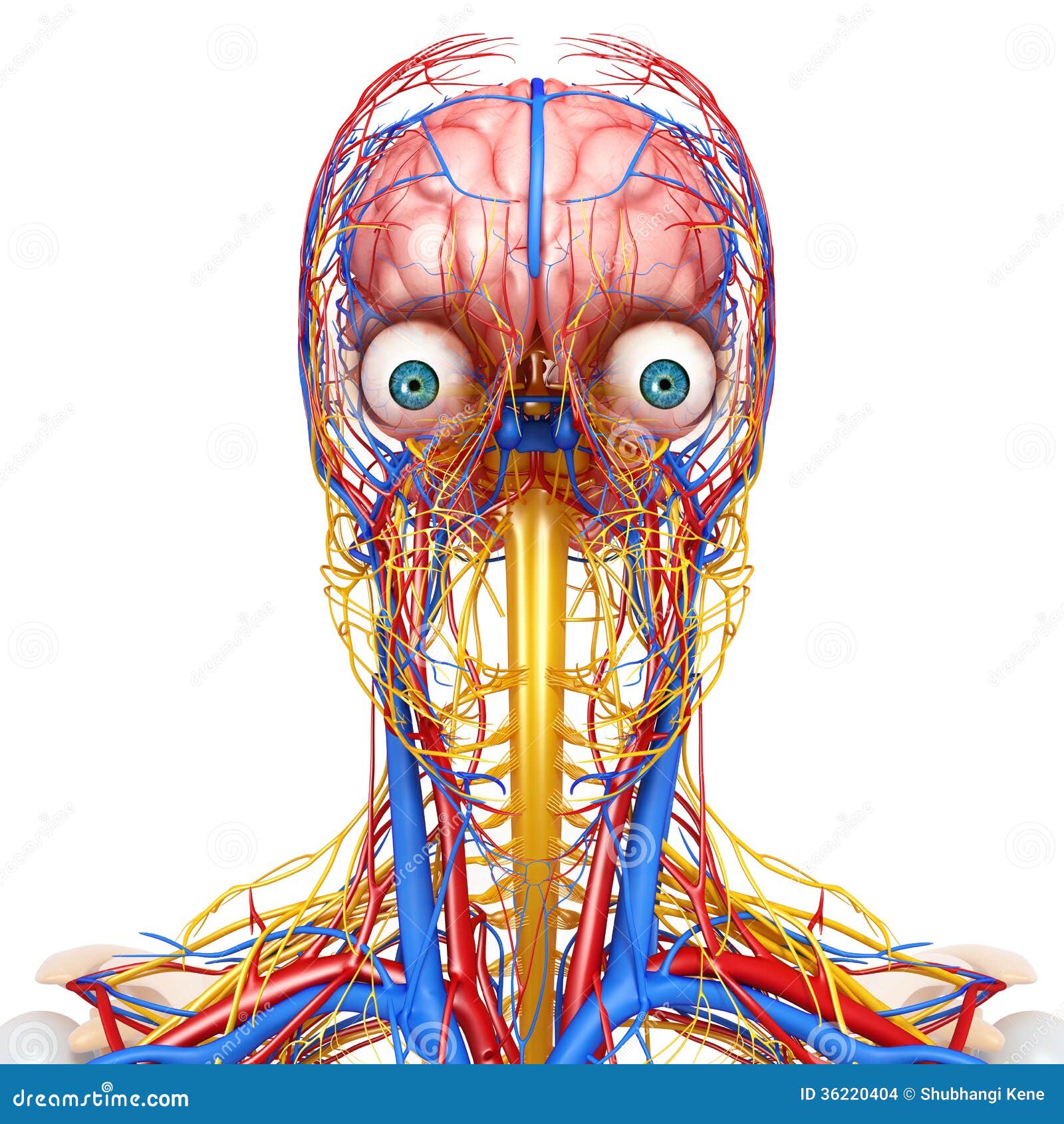
Circulatory and Nervous System of Head Stock Illustration ...
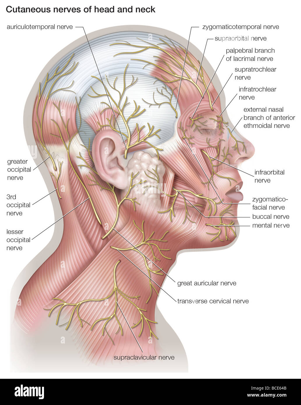
Diagram of the cutaneous nerves of the head and neck Stock ...
Superficial nerves of the face and scalp: Anatomy | Kenhub

Cervical Spine Anatomy (Neck)
8,361 Human Head Diagram Stock Photos, Pictures & Royalty ...

Occipital Neuralgia
Superficial nerves of the face and scalp: Anatomy | Kenhub
The 12 Cranial Nerves and their Functions | Medical Library
0 Response to "39 nerves in the head diagram"
Post a Comment