37 diagram of synovial joints
› metacarpal-bonesMetacarpals: Definition, Location, Anatomy, Function, Diagram The joints between the metacarpal and carpal bones, these are all plane synovial joints, except the thumb as it is a saddle joint (another form of synovial joint) [8]. The five metacarpals form prominent articulations on their base or proximal end, with one or more of the four distal carpal bones [4] : Synovial Joints - Anatomy and Physiology synovial joint at which the convex surface of one bone articulates with the concave surface of a second bone; includes the elbow, knee, ankle, and interphalangeal joints; functionally classified as a uniaxial joint. intracapsular ligament. ligament that is located within the articular capsule of a synovial joint.
study.com › academy › lessonJoints: Structure and Functions - Video & Lesson Transcript ... Aug 24, 2021 · Learn about the structure of joints, the function of joint capsules, synovial membrane, bursa, meniscus, and the importance of synovial fluid in reducing friction between bones. Updated: 08/24/2021
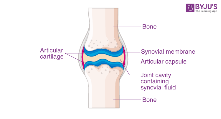
Diagram of synovial joints
Synovial Joint - SmartDraw Synovial Joint. Create healthcare diagrams like this example called Synovial Joint in minutes with SmartDraw. SmartDraw includes 1000s of professional healthcare and anatomy chart templates that you can modify and make your own. 26/37 EXAMPLES. Synovial Joints Teaching Resources | Teachers Pay Teachers This is a comprehensive test that includes vocabulary terms, a diagram of the composition of a bone, diagram of a synovial joint, and diagram of the elementary skeleton labeling. This test is designed to pair with the coordinating vocabulary terms and activities in this shop. This test was designed for 4th, 5th, 6th, or 7th grade science ... Joints of the skeletal system - Skeletal system - Edexcel ... In synovial joints, the ends of the bones are covered with cartilage (called articular cartilage) which cushions the joint and prevents friction and wear and tear between the bone ends. Cartilage ...
Diagram of synovial joints. PDF Joints Classification of Joints • 1. According to the type of tissue at the joint: • a) Fibrous joint -- uses fibrous connective tissue to articulate bones. • b) Cartilaginous joint-- uses hyaline cartilage and/or fibro- cartilage to articulate bones. • c) Synovial joint --uses auricular cartilage, synovial membrane, joint capsule, and ligaments to articulate bones. Types of Synovial Joints | Biology for Majors II Synovial joints are further classified into six different categories on the basis of the shape and structure of the joint. The shape of the joint affects the type of movement permitted by the joint (Figure 1). These joints can be described as planar, hinge, pivot, condyloid, saddle, or ball-and-socket joints. Figure 1. Synovial Joint Anatomy in Animal - Definition, Types and ... The synovial joint is a moveable or true joint in an animal's body. Hi there, do you want to learn synovial joint anatomy in animals? Fine, in this article, I will describe the synovial joint structure with a labeled diagram. I will also describe different types of synovial joints in animals. After reading this article, you will know the ... Joint: synovial - MyDr.com.au Synovial joints may also become inflamed, called arthritis. There are more than 100 different types of arthritis, arising from problems in different parts of the joint. For example in osteoarthritis, the cartilage becomes worn , and in rheumatoid arthritis the body's immune system attacks the synovial membrane.
Labelled Diagram Of Synovial Joint - schematron.org Synovial joints allow for smooth movements between the adjacent bones. This diagram shows the location of the bursae which are fluid filled sacs in a bone. The basic structure of a synovial joint is shown in the diagram below. The main parts of synovial joints are labelled on the synovial joint diagram. The Joints of the Body - Quiz 1 - Free Anatomy Quiz Which type of synovial joint are the carpometacarpal joints (label C in the image)? Which of the following is an example of a synovial plane joint shown in the image above? What sort of synovial joint is the hip joint (the acetabulofemoral joint; label E in the image)? Which type of joints are held together by hyaline cartilage or fibrocartilage? byjus.com › biology › saddle-jointsAnatomy and Physiology Of Saddle Joints - An Overview A synovial joint is one among the three types of joints, which are classified based on their structure and is one of the most common types of joints in the human body. Synovial joints are more flexible and movable joints, which perform a wide range of locomotion , such as walking, running, typing and more. The Six Types of Synovial Joints: Examples & Definition ... Next, let's focus on hinge joints, shown as letter B on the diagram. Hinge joints are the synovial joint type referred to in our introductory section. These joints can be found between your upper and lower arm bones, otherwise called your elbow, as well as your ankles, fingers, toes, and knees. Hinge joints operate just like the hinges on a door.
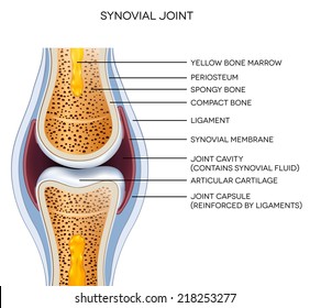
Structure of a synovial joint pdf - Australia Tutorials ... The main parts of synovial joints are labelled on the synovial joint diagram and described in the table below. A, The egg-shaped ovoid surface represents a characteristic of most synovial joints of the body (e.g., hip joint, radiocarpal joint, knee joint, metacarpophalangeal joint). The diagram shows only the convex member of the joint. › biology › ellipsoid-jointsEllipsoid Joints - Meaning, Types, Features, and FAQs Synovial joints are more complex than the other types of joint and their structural components include. Synovial fluid . Articular capsule . Articular cartilage . Reinforcing ligaments . Joint cavity or capsules . Types of Synovial Joints . Synovial joints are also called diarthrosis joints and almost all the joints present in our body are ... Diagram Of Synovial Joint - Elcacerolazo Synovial Joint Diagram Labeled Anatomy Chart With Two Bones Synovial joints are characterized by the presence of a joint cavity. It consists of two layers. Fine in this article I will describe the synovial joint structure with a labeled diagram. Allows movement in only one plane. Tough Fibrous tissue surrounds synovial joints. Structures of a Synovial Joint - Capsule - Ligaments ... A synovial joint is characterised by the presence of a fluid-filled joint cavity contained within a fibrous capsule. It is the most common type of joint found in the human body, and contains several structures which are not seen in fibrous or cartilaginous joints.. In this article we shall look at the anatomy of a synovial joint - the joint capsule, neurovascular structures and clinical ...
Draw A Labelled Diagram Of A Synovial Joint. Give Examples ...
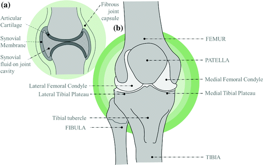
(a) A Synovial Joint; (b) Types of Synovial Joints. Image ... Development of a Low-Cost and Easy-to-Use Wearable Knee Joint Monitoring System. Thesis. Full-text available. Apr 2020. Abu Ilius Faisal. The loss of mobility among the elderly has become a ...

Synovial joint diagram. Labeled anatomy chart with two bones ...
The Six Types of Synovial Joints: Examples & Definition ... Next, let's focus on hinge joints, shown as letter B on the diagram. Hinge joints are the synovial joint type referred to in our introductory section. These joints can be found between your upper ...
Joints
open.oregonstate.education › 9-4-synovial-joints9.4 Synovial Joints – Anatomy & Physiology Synovial joints are subdivided based on the shapes of the articulating surfaces of the bones that form each joint. The six types of synovial joints are pivot, hinge, condyloid, saddle, plane, and ball-and socket-joints (Figure 9.4.3).

Stock vektor „Synovial Joint Anatomy Joint Capsule Synovial ...
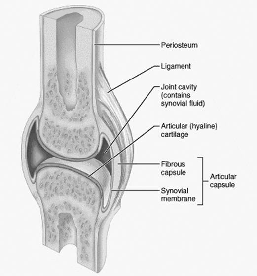
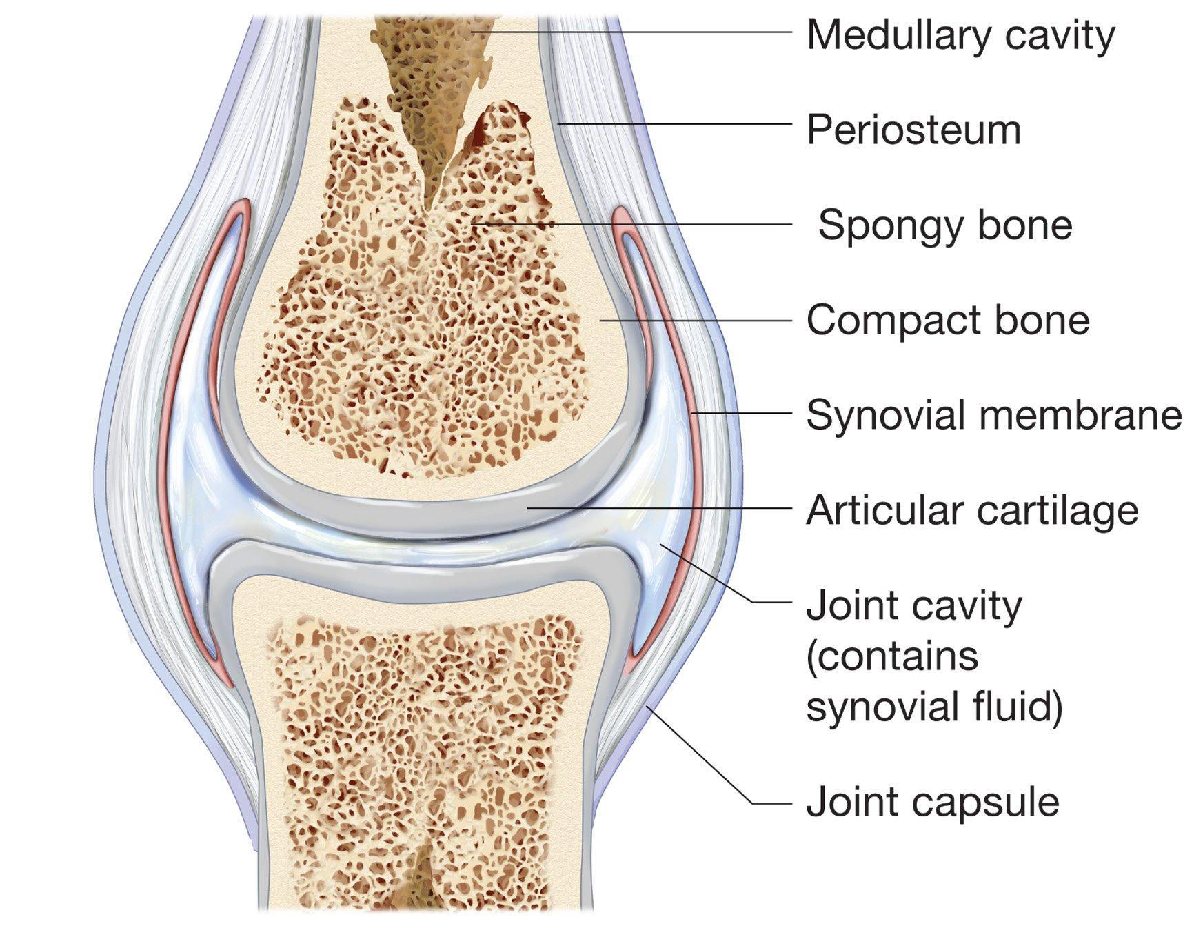
3. What characteristics do all joints have in common ... 3. What characteristics do all joints have in common? 4. Label the diagram of a typical synovial joint using the terms provided in the key and the appropriate leader lines. Key: a. articular capsule b. articular cartilage c. fibrous layer d. joint cavity e. ligament f. periosteum 9. synovial membrane. Question: 3.
What Is a Synovial Joint?
Joints and Ligaments | Learn Skeleton Anatomy Synovial joints are often supported and reinforced by surrounding ligaments, which limit movement to prevent injury. There are six types of synovial joints: (1) Gliding joints move against each other on a single plane. Major gliding joints include the intervertebral joints and the bones of the wrists and ankles.
BTEC Revision Guide Skeletal System
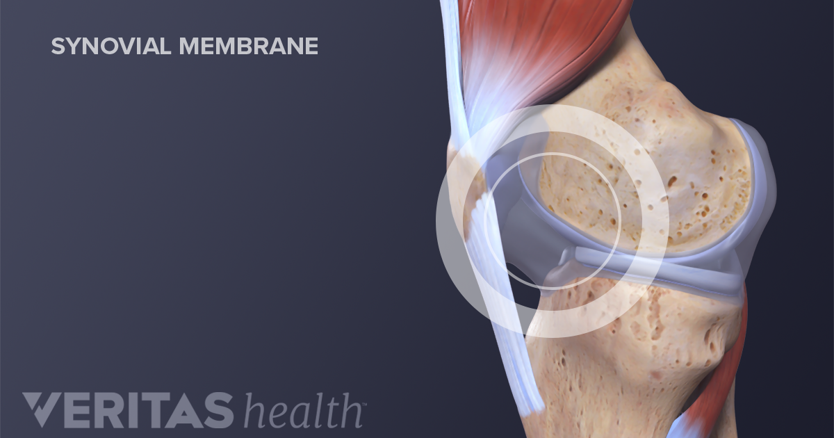
Synovial Joints | Boundless Anatomy and Physiology A synovial membrane (or synovium) is the soft tissue found between the articular capsule (joint capsule) and the joint cavity of synovial joints. Synovial fluid is the clear, viscid, lubricating fluid secreted by synovial membranes. The morphology of synovial membranes may vary, but it often consists of two layers.
Different Types of Joints in Body | Bone and Spine
Types of Synovial Joints label Diagram | Quizlet Types of Synovial Joints label. STUDY. Learn. Flashcards. Write. Spell. Test. PLAY. Match. Gravity. Created by. elizabethsisley. Terms in this set (6) Pivot Joint. allow for rotation around an axis, such as between the first and second cervical vertebrae, which allows for side-to-side rotation of the head. Hinge Joint.

34 Synovial fluid Vector Images, Synovial fluid Illustrations ...
Solved 6. all are freely movable or diarthrotic 2. Label ... 4. Which structure in the synovial joint; Question: 6. all are freely movable or diarthrotic 2. Label the diagram of a typical synovial joint using the terms provided in the key and the Key: a. articular capsule b. articular cartilage C. fibrous layer d. joint cavity e. ligament f. periosteum g. synovial membrane 3.

Anatomy: Synovial Joint Diagram Diagram | Quizlet
Bones Joints and Cartilage Notes: Diagrams & Illustrations ... CARTILAGINOUS JOINTS Hyaline cartilage connects bones, stretches to allow some movement Synchondrosis: costochondral joint, where cartilage attaches rib to sternum; growth plates between bone diaphysis, epiphysis Symphysis: symphysis pubis in pelvic bone (fibrous cartilage) ↑ strength, ↓ flexibility SYNOVIAL JOINTS Joint capsule connects ...
How to describe the composition of the essential structure ...
PDF Joints Of The Body Worksheets Answer Key - Weebly Extremely stable joint. 9. Figure 8.3 shows diagrams of two synovial joints. Identify each joint by inserting the name of the joint in the blank below each diagram. Then select different colors, and use them to color the coding circles and the structures that are present in the diagrams. Finally, add labels and leadeç lines an the Labels C 3 p/s)

Do all joints in the human body contain synovial fluid? Are ...
Labelled Diagram Of Synovial Joint - Wiring Diagrams Labelled Diagram Of Synovial Joint. A synovial joint is a connection between two bones consisting of a cartilage lined As seen in the above picture, the most powerful bite in the world gets its. A synovial joint or diarthrosis occurs at articulating bones to allow movement. fibrous connective tissue found in various parts of the body such as ...

synovial joint. Easy pic for patients to understand and you ...
Synovial Joint Diagram Label - schematron.org The structure of a synovial joint is demonstrated by a diagram in which the articulating bones are surrounded by the articular capsule, which comprises an exterior fibrous capsule and an interior synovial membrane. Start studying label the synovial joint. Learn vocabulary, terms, and more with flashcards, games, and other study tools.

Synovial Joint Diagram Labeled Stock Vector - Illustration of ...
Synovial Joints Anatomy Diagram | Quizlet The only 2 synovial joints that aren't diarthrotic. carpals and tarsals. Functional classification of carpals and tarsals. amphiarthrosis. membrane continuous from bone to bone outside the articular capsule. periosteum. fibrous capsule lined by synovial membrane. articular capsule.

Synovial fluid - Wikipedia
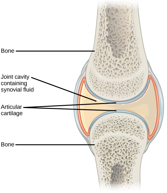
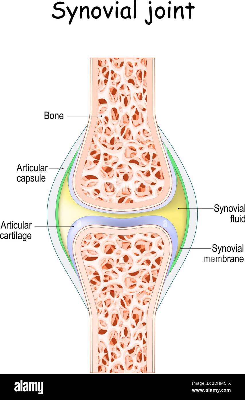
courses.lumenlearning.com › boundless-ap › chapterClassification of Joints | Boundless Anatomy and Physiology Synovial Joint: This diagram of a synovial joint delineates the articular cartilage, articular capsule, bone, synovial membrane, and joint cavity containing synovial fluid. Synovial Joints This is the most common and movable joint type in the body.

Describe typical synovial joint with a neat labelled diagram ...
[SOLVED] Draw a labelled diagram of a synovial joint. Give ... Draw a labelled diagram of a synovial joint. Give one example each for a hinge joint, a pivot joint, axial skeleton and appendicular skeleton. Medium. Open in App. Solution. Verified by Toppr. Examples of: 1. Hinge joint: Allows movement in only one plane. Elbow joint and knee joint. 2. Pivot joint: Primary movement is a rotation.

Synovial Joint Anatomy in Animal - Definition, Types and ...
byjus.com › biology › synovial-jointsAnatomy and Physiology Of Synovial Joints - An Overview Synovial Joints. Joints can be simply defined as articulations of bones, which functions by providing shape to the skeleton system, protects bones by holding them together securely and also helps in movement. Based on structure and functions, joints have been further classified into different types. A synovial joint is one among the three types ...

Download synovial images for free
Joints of the skeletal system - Skeletal system - Edexcel ... In synovial joints, the ends of the bones are covered with cartilage (called articular cartilage) which cushions the joint and prevents friction and wear and tear between the bone ends. Cartilage ...
Synovial Knee Joint | SpringerLink
Synovial Joints Teaching Resources | Teachers Pay Teachers This is a comprehensive test that includes vocabulary terms, a diagram of the composition of a bone, diagram of a synovial joint, and diagram of the elementary skeleton labeling. This test is designed to pair with the coordinating vocabulary terms and activities in this shop. This test was designed for 4th, 5th, 6th, or 7th grade science ...

The structure of a synovial joint. | Synovial joint, Joints ...
Synovial Joint - SmartDraw Synovial Joint. Create healthcare diagrams like this example called Synovial Joint in minutes with SmartDraw. SmartDraw includes 1000s of professional healthcare and anatomy chart templates that you can modify and make your own. 26/37 EXAMPLES.

Structure of synovial joint

Synovial joints(in Hindi)
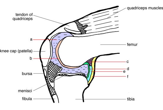
![A typical synovial joint (the knee joint) [19]. | Download ...](https://www.researchgate.net/profile/Houssein-Lamine-2/publication/339686957/figure/fig4/AS:865361939402752@1583329772113/A-typical-synovial-joint-the-knee-joint-19.jpg)
A typical synovial joint (the knee joint) [19]. | Download ...
Synovial joint activity
What is the function of the synovial membrane of a Synovial ...
19.3 Joints and Skeletal Movement – Concepts of Biology – 1st ...

Synovial Joints | Anatomy and Physiology I
Synovial joint | Radiology Case | Radiopaedia.org
Synovial joints Images, Stock Photos & Vectors | Shutterstock
Synovial joint - Teaching resources
Synovial joint anatomy. joint capsule with synovial fluid and ...

Synovial joint - Wikipedia
Structure and function of synovial joints – HSC PDHPE

Synovial joint structure stock vector. Illustration of ...

Synovial Joint - an overview | ScienceDirect Topics
Joints

Types of Synovial Joints | Biology for Majors II

Structures of a Synovial Joint - Capsule - Ligaments ...
0 Response to "37 diagram of synovial joints"
Post a Comment