37 sheep brain diagram labeled
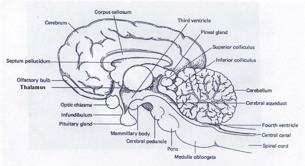
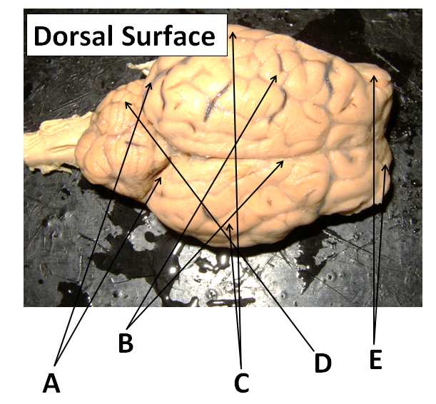
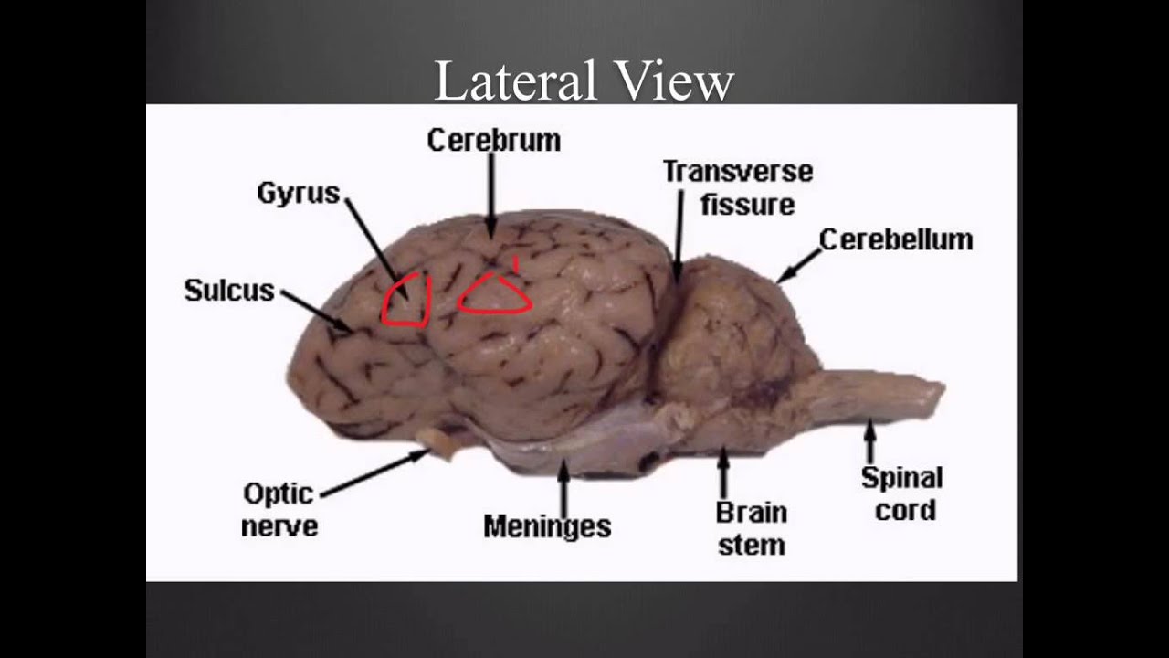
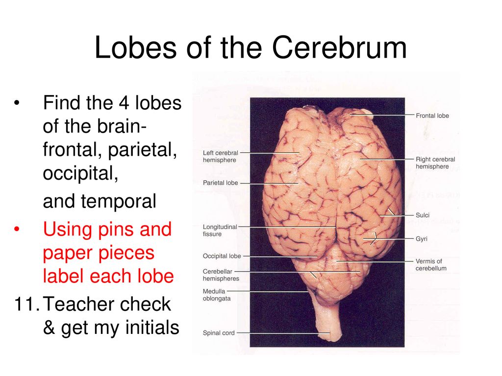
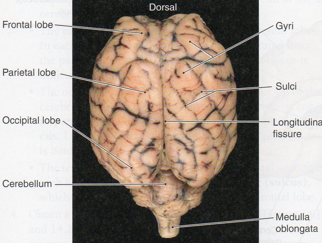
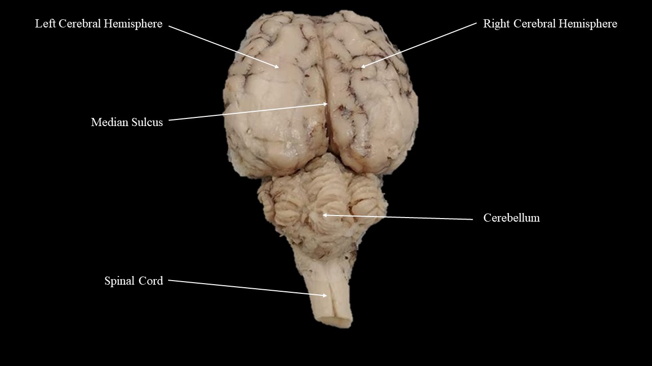
Sheep Brain Dissection Lab The lobes of the brain are visible, as well as the transverse fissure, which separates the cerebrum from the cerebellum. The convolutions of the brain are also visible as bumps (gyri) and grooves (sulci). Use the diagram below to help you locate these items. Dorsal View of the Sheep Brain . 8. PDF Sheep Brain Midsagittal Section - Dr. Scott Croes' Website 5 3 11 6 22 16 18 1. Gray Matter 2. White Matter 3. Corpus Callosum 4. Lateral Ventricle 5. Caudate Nucleus 6. Septum Pellucidum 7. Fornix 8.
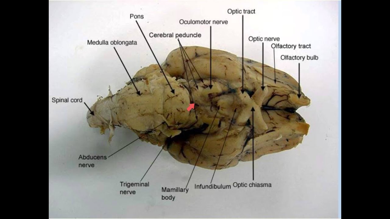
PDF Lab: Sheep Brain Dissection - Mrs. Moretz's Science Site to anatomy studies. See for yourself what the . cerebrum, cerebellum, spinal cord, gray matter, white matter, and other parts of the brain look like! Observation: External Anatomy . 1. You'll need a . preserved sheep brain. for the dissection. Set the brain down so the flatter side, with the white . spinal cord. at one end, rests on the ...

Sheep brain diagram labeled
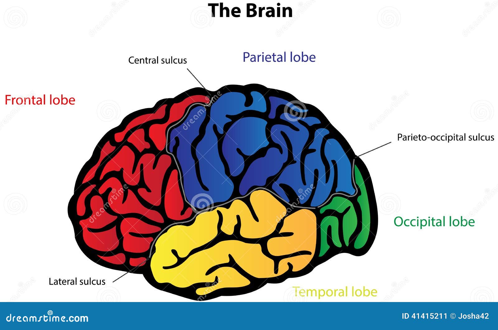
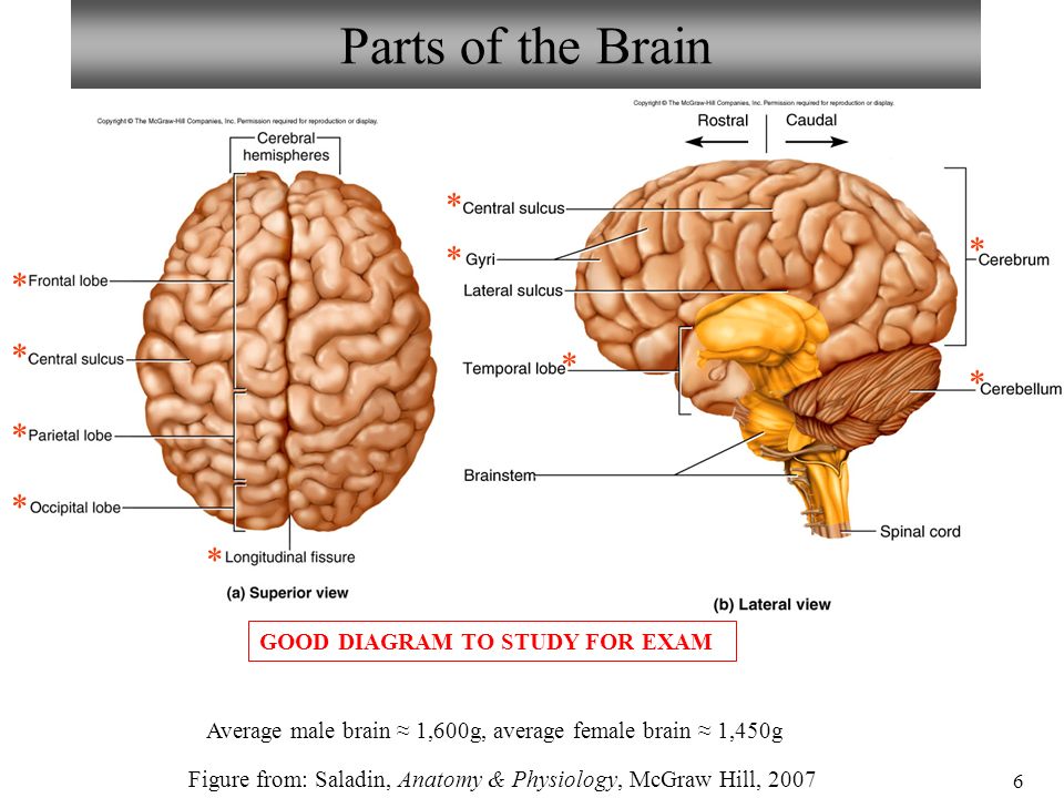
Brain Lobes Diagram Labeled - Studying Diagrams With more related things such sheep brain diagram labeled brain nervous system worksheet and blank heart diagram. The diagram of the brain is useful for both Class 10 and 12. THE LOBES Occipital lobe Lower back of the brain. Sheep Brain Dissection Project Guide | HST Learning Center Use the labeled picture to identify the corpus callosum, medulla, pons, midbrain, and the place where the pituitary gland attaches to the brain. (In many preserved specimens the pituitary gland is no longer present. It is not pictured.) Use your fingers or a teasing needle to gently probe the parts and see how they are connected to each other. Sheep Brain Anatomy #2 Diagram - Quizlet Sheep Brain Anatomy #2. STUDY. PLAY. cerebellum. posterior part of the brain that coordinates muscle movements and maintains balance. temporal lobe. interpretation and integration of speech and sound. parietal lobe. interpretation and integration of sensory stimuli. frontal lobe.
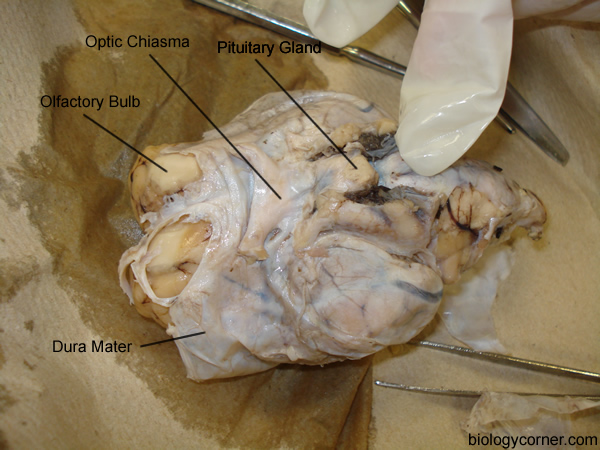
Sheep brain diagram labeled. Sheep brain labeling Quiz - PurposeGames.com This online quiz is called Sheep brain labeling. This game is part of a tournament. You need to be a group member to play the tournament Sheep Brain Anatomy Quiz - ProProfs Quiz Sheep Brain Anatomy Quiz. Sheep are wonderful and cute. The brain is an interesting organ. It helps with cognition and memory. Almost all the basic task In the body is commanded by the Brain. It is the control center of the body which regulates and control the process crucial for survival Are you interested in learning more about the brain of ... Sheep Brain Dissection labeled 2 Diagram - Quizlet Start studying Sheep Brain Dissection labeled 2. Learn vocabulary, terms, and more with flashcards, games, and other study tools. Sheep Brain Dissection with Labeled Images The sheep brain is exposed and each of the structures are labeled and described in a sequential manner, in the same way that a real dissection would occur. Sheep Brain Dissection with Labeled Images Sheep Brain Dissection 1. The sheep brain is enclosed in a tough outer covering called the dura mater.
Sheep Brain Diagram - Diagram Sketch Sheep Brain Diagram. angelo on October 9, 2021. Image Result For Sheep Brain Labeled Brain Diagram Human Brain Diagram Brain Anatomy. Sheep Brain Dissection Guide With Pictures Worksheets Nervous System Anatomy Brain Anatomy Dissection. Sheep Brain External View Labeled Anatomia Veterinaria Anatomia Veterinaria. labeled brain | Brain anatomy, Human anatomy and ... Sheep Brain Dissection Guide. In this lab guide, students are given instruction on how to remove the dura mater, and locate the main structures of the external and internal brain. Becky Aguiar. School-Science. Human Brain Anatomy. Human Anatomy And Physiology. Brain Science. Medical Science. Nervous System Anatomy. Sheep Brain Labeled Diagram - Diagram Sketch Sheep Brain Labeled Diagram. angelo on January 2, 2022. Horse Brain 2 Brain Anatomy Brain Diagram Nervous System Anatomy. Labeled Diagram Of Brain Midsagittal View Diigo Groups Brain Anatomy Anatomy And Physiology Human Brain Anatomy. Sheep Brain Dissection Guide With Pictures Worksheets Nervous System Anatomy Brain Anatomy Dissection. Image ... PDF Sheep Brain Dissection - Administration Sheep Brain Dissection The purpose of this exercise is to introduce you to the mammalian brain; you will use a sheep's brain. While the sheep brain differs from the human brain in many details, they both have the same basic anatomy, and, it is larger than the rat brain. Work in teams of four students.
A virtual sheep brain dissection guides anatomy studies ... Sheep Brain Dissection with Labeled Images The sheep brain is exposed and each of the structures are labeled and described in a sequential manner, in the same way that a real dissection would occur. A Amanda Huss Anatomy Brain Anatomy Human Anatomy And Physiology Medical Anatomy Body Anatomy Skull Anatomy Nervous System Anatomy Brain Models Label Sheep Brain Diagram | Quizlet Label Sheep Brain. STUDY. Learn. Flashcards. Write. Spell. Test. PLAY. Match. Gravity. Created by. telaneyn. Terms in this set (11) Cerebellum. the "little brain" at the rear of the brainstem; functions include processing sensory input and coordinating movement output and balance. Occipital lobe. Brain Dissection Diagram - Diagram Sketch Sheep Brain Dissection Lab Companion In 2021 Brain Anatomy Anatomy And Physiology Brain. Labeled Brain Model Bing Images Practical And Final Nerve Anatomy Brain Models Nervous System Anatomy. Labeled Diagram Of Brain Midsagittal View Diigo Groups Brain Anatomy Anatomy And Physiology Human Brain Anatomy. Sheep Brain Dissection Project Guide Hst ... Sheep Brain Diagram Unlabeled - Diagram Sketch Sheep Brain Diagram Unlabeled. Parts Of The Brain Brain Diagram Function Diagram Human Brain Diagram. Sheep Brain Label Brain Diagram Dissection Brain Anatomy. Pin By Melissa On Brain Anatomy In 2021 Human Brain Diagram Brain Diagram Basic Anatomy And Physiology. Great For Grade 5 Science Brain Craft The Brain For Kids Brain Anatomy.
Sheep brain images | Lab | Amherst College
PDF Neuroanatomy: Dissection of the Sheep Brain Examine the sheep brain with the membranes intact. You should be able to identify and use the following directional terms: Anterior / Posteriorfront / back Rostral / Caudal towards the beak / towards the tail Medial / Lateral towards the middle / towards the side Dorsal / Ventral top / bottom (on the CNS of a quadruped)

Pretty good picture of the sheep brain labeled. | Basic ...
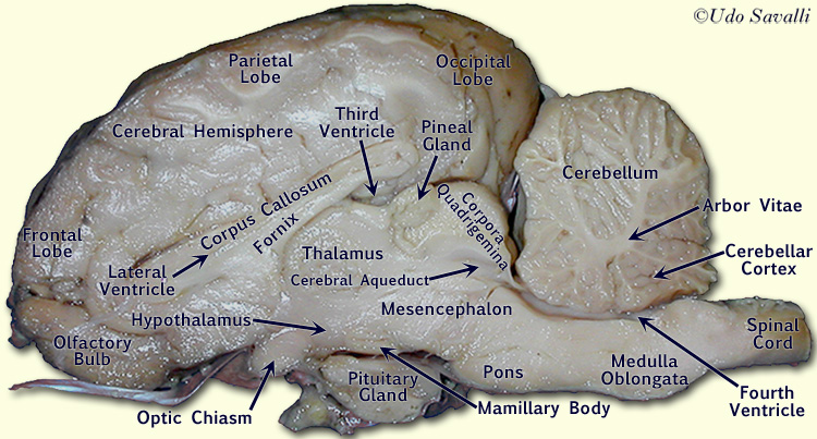
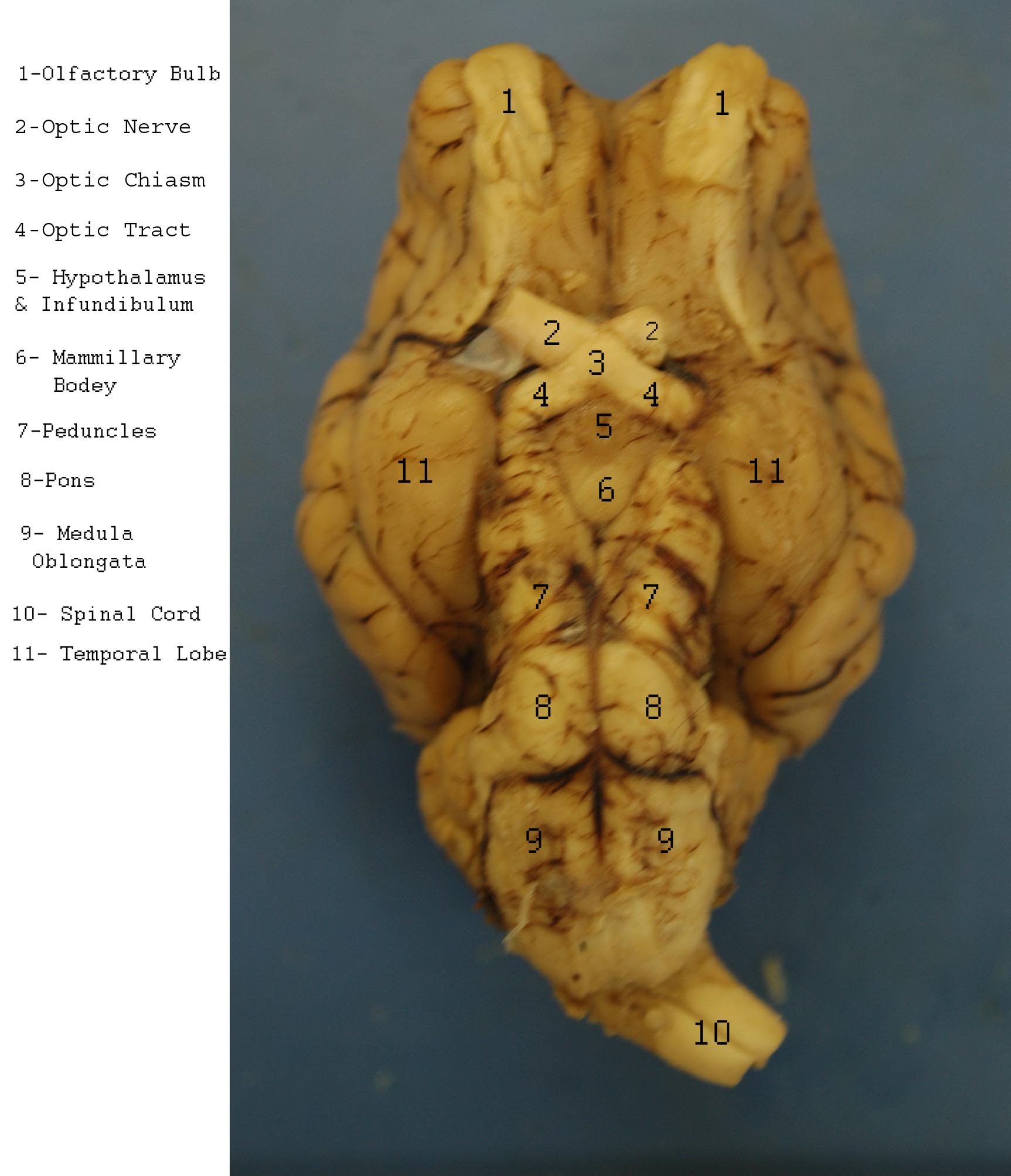
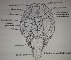
Sheep brain dissection | Human Anatomy and Physiology Lab ... The most prominent structure visible on the ventral side of the sheep brain is half of the optic chiasma, which is where the two optic nerves cross over each other and form an "X" shape. You will only see half the structure. Find the optic chiasma half on your brain. You may have removed the optic removed the chiasma with the dura mater.

Sheep Brain Images
DOC Sheep Brain Anatomy Lab Manual - amherst.edu Sheep Brain Anatomy Lab Manual. Based on original material by R. N. Leaton, Dartmouth College. Contributors to this version: Al Sorenson, Lisa Raskin, Sarah Turgeon, Steve George, and JP Baird. I. Introduction. The brain of the sheep is useful for study because its anatomy is similar to human brain anatomy. Although exact proportions (and names ...

Sheep Brain
Sheep Brain Anatomy and Function Flashcards - Cram.com Study Flashcards On Sheep Brain Anatomy and Function at Cram.com. Quickly memorize the terms, phrases and much more. Cram.com makes it easy to get the grade you want!
Physiological Psychology
PDF Distance Learning Program Anatomy of the Human Brain/Sheep ... function, and pathology. Those students participating in Sheep Brain Dissections will have the opportunity to dissect and compare anatomical structures. At the end of this document, you will find anatomical diagrams, vocabulary review, and pre/post tests for your students. The following topics will be covered: 1.
Bio211 Laboratories 8 & 9 Brain/Cranial Nerves Spinal Cord ...
PDF DISSECTION OF THE SHEEP'S BRAIN - Hanover College DISSECTION OF THE SHEEP'S BRAIN Introduction The purpose of the sheep brain dissection is to familiarize you with the three-dimensional structure of the brain and teach you one of the great methods of studying the brain: looking at its structure. One of the great truths of studying biology is the saying that "anatomy precedes physiology".
The brain | Annette's Vet Student Info
Sheep Brain Label | Brain diagram, Dissection, Brain anatomy Sheep Brain Label A drawing of the brain with the parts unlabeled. Students can practice naming the parts of the brain, then check their answers with the provided key. Biologycorner 17kfollowers More information unlabeled brain Find this Pin and more on A&Pby Dijana Kovacevic. Brain Gym For Kids Human Brain Diagram Brain Anatomy And Function

Sheep brain dissection | Human Anatomy and Physiology Lab ...
Sheep Brain Quiz - PurposeGames.com How is Your Memory? 11p Text Game. METRIC SYSTEM - the basics 10p Image Quiz. Countries of the European Union (by shape) 27p Image Quiz. 13 Colonies Quiz 13p Image Quiz. Sightseeing the US 28p Image Quiz. Math Theorems and Constants 14p Image Quiz. PG Lightning Game: Speed counting 20p Shape Quiz.
Sheep Brain Neuroanatomy Online Self-Test | KPU.ca - Kwantlen ...
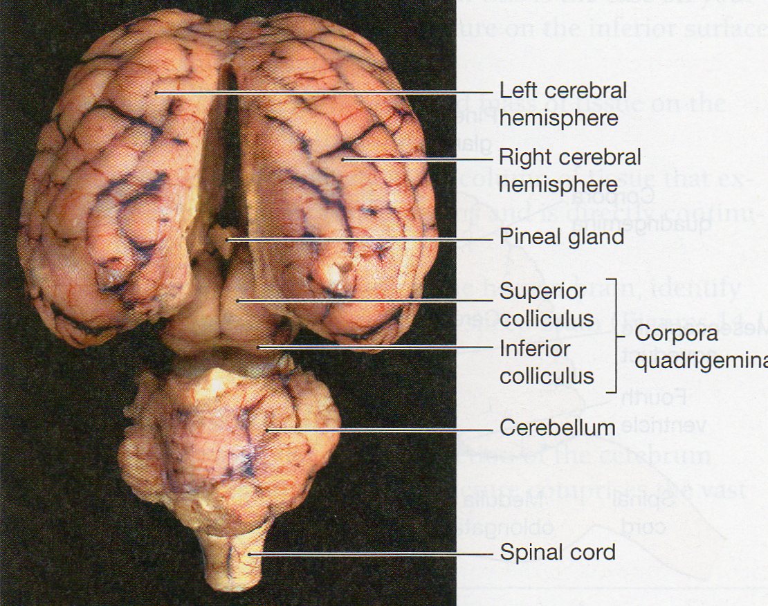
Sheep Brain - Veterinary Anatomy Website Home Page Dorsal view of sheep brain with the cerebellum and caudal cerebrum removed. The rostral colliculus (large arrow label) and the caudal colliculus (small arrow label) together form the tectum of the midbrain.. Also labeled are the pineal body (green), the caudate nucleus (1), the floor of the fourth ventricle (white and pink) and cerebellar peduncles (blue = rostral, red = middle, and yellow ...
Sheep Brain Dissection with Labeled Images
Sheep Brain Dissection labeled Diagram | Quizlet Start studying Sheep Brain Dissection labeled. Learn vocabulary, terms, and more with flashcards, games, and other study tools.
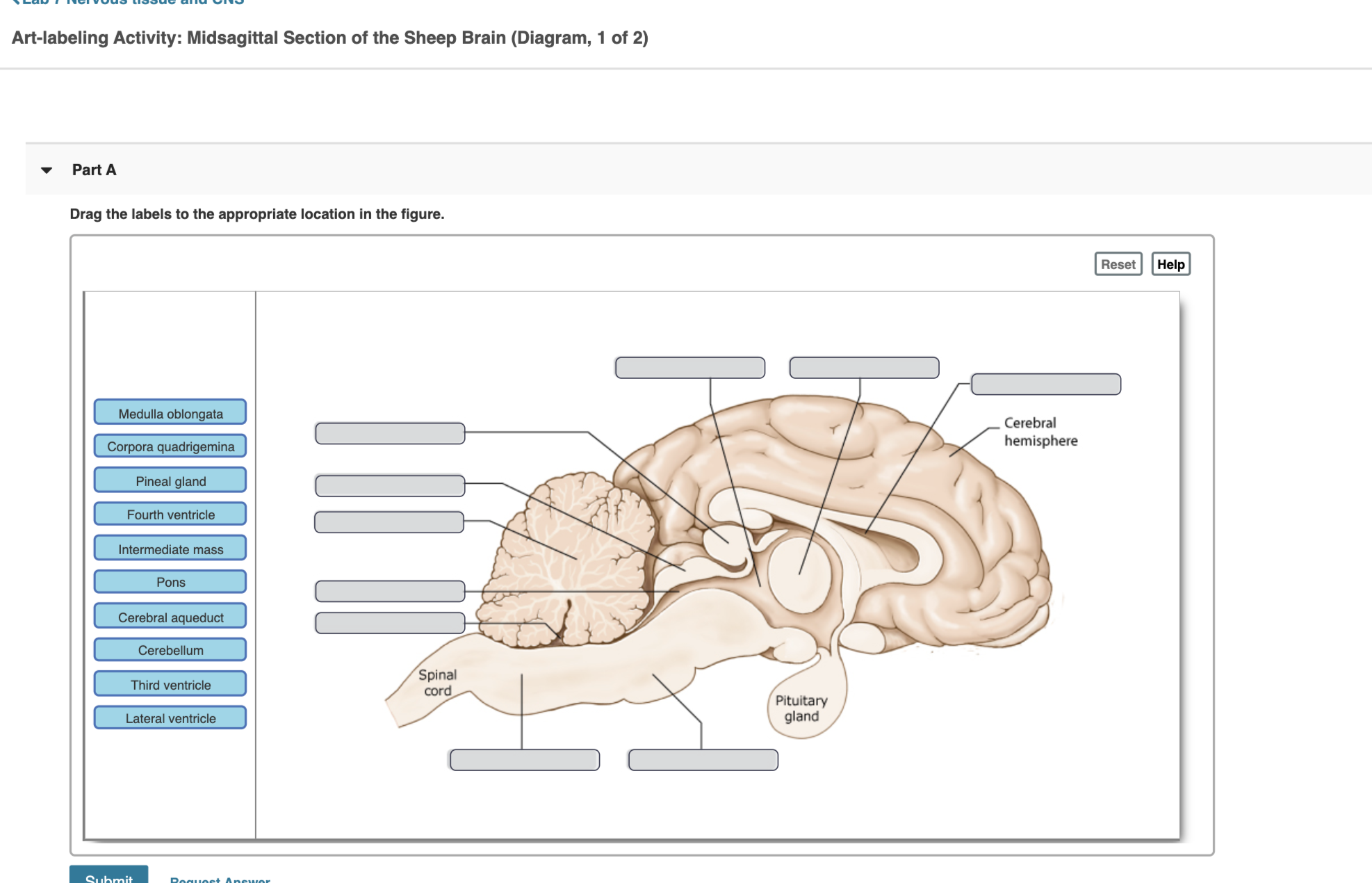
Solved Art-labeling Activity: Midsagittal Section of the ...
Sheep Brain Label - The Biology Corner Label theBrain of the Sheep. Publisher: Biologycorner.com; follow on Google+ This work is licensed under a Creative Commons Attribution-NonCommercial 3.0 Unported License. Brain Label Answer Key. Image adapted from a photograph of the sheep brain. ...
BIO201-Sheep Brain
Sheep Brain Anatomy #2 Diagram - Quizlet Sheep Brain Anatomy #2. STUDY. PLAY. cerebellum. posterior part of the brain that coordinates muscle movements and maintains balance. temporal lobe. interpretation and integration of speech and sound. parietal lobe. interpretation and integration of sensory stimuli. frontal lobe.
Pin von Iris Yeh auf Final anatomy
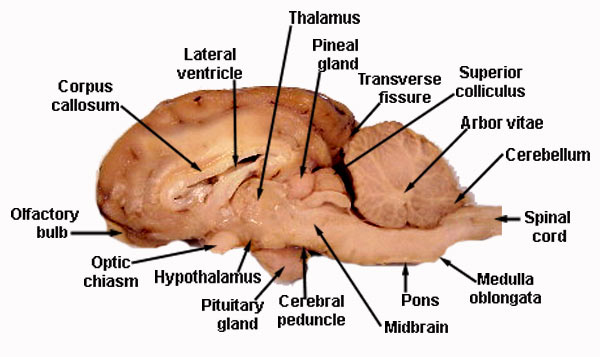
Sheep Brain Dissection Project Guide | HST Learning Center Use the labeled picture to identify the corpus callosum, medulla, pons, midbrain, and the place where the pituitary gland attaches to the brain. (In many preserved specimens the pituitary gland is no longer present. It is not pictured.) Use your fingers or a teasing needle to gently probe the parts and see how they are connected to each other.
Sheep Brain Dissection Guide
Brain Lobes Diagram Labeled - Studying Diagrams With more related things such sheep brain diagram labeled brain nervous system worksheet and blank heart diagram. The diagram of the brain is useful for both Class 10 and 12. THE LOBES Occipital lobe Lower back of the brain.
Sheep Brain Dissection Report

Sheep brain sagittal section medial view - www.anatomynote ...

label median section of sheep brain Diagram | Quizlet
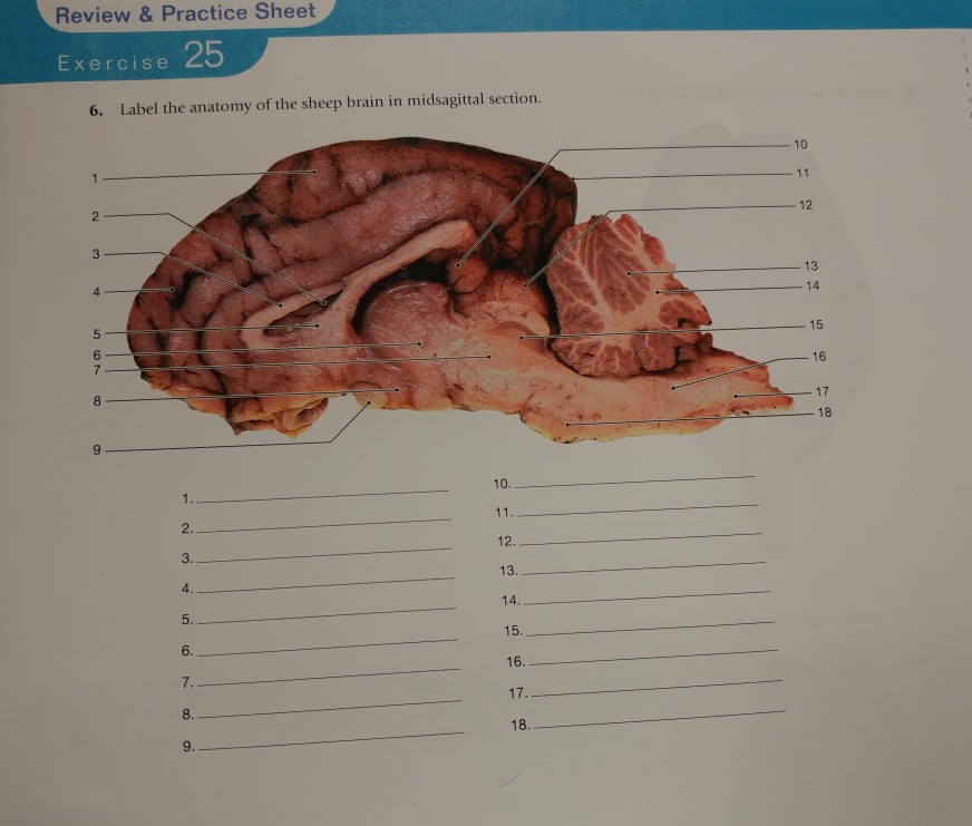
Solved Review & Practice Sheet Exercise 6. Label the anatomy ...
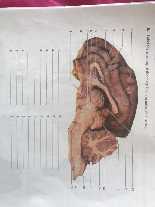
Solved 6. Label the anatomy of the sheep brain in | Chegg.com
Sheep Brain Dissection Guide - ppt download
Sheep Neuroanatomy Lab- Labeling Worksheet Figure 1: Dorsal view

Sheep Brain Dissection labeled Diagram | Quizlet
Medical Detectives Lesson 28
Brain Anatomy Labeled Diagram Stock Vector - Illustration of ...

Sheep Brain
Physiological Psychology

Sheep Brain Quiz

Sheep Brain
BIO201-Sheep Brain

BIO201-Sheep Brain
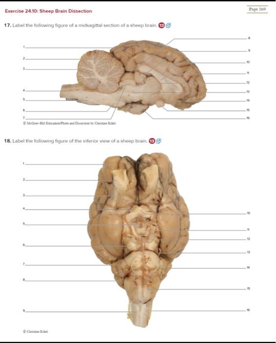
Solved Exercise 24.10: Sheep Brain Dissection Page 509 ...
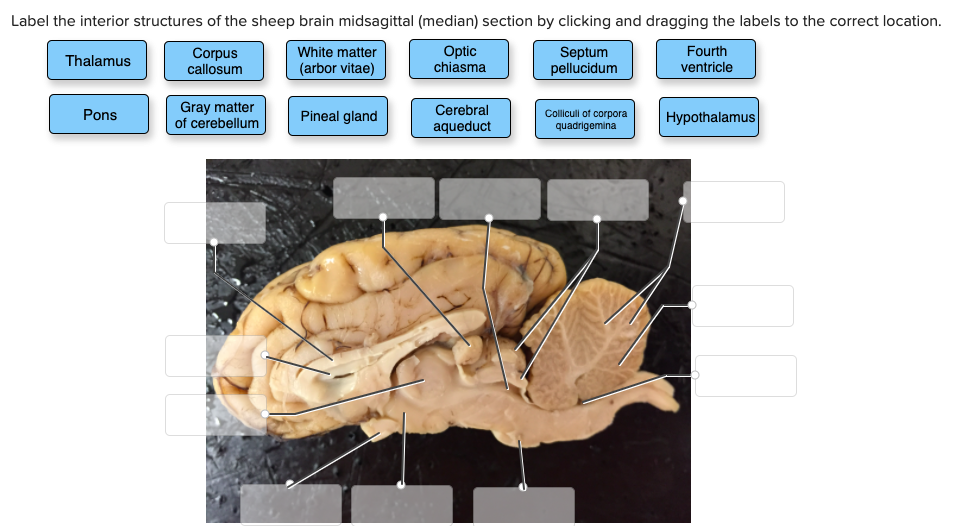
Solved Label the interior structures of the sheep brain ...
Index of /files/OCC_VIDEO/upload/Faculty_Resources/acamilo ...
Neuron/Spinal Cord Histology Brain Anatomy Sheep Brain ...
3.2: Sheep brain - Medicine LibreTexts
Physiological Psychology
Lab Exam 3: Anatomy of Sheep Brain; Histology Flashcards ...
0 Response to "37 sheep brain diagram labeled"
Post a Comment