38 meiotic division beads diagram
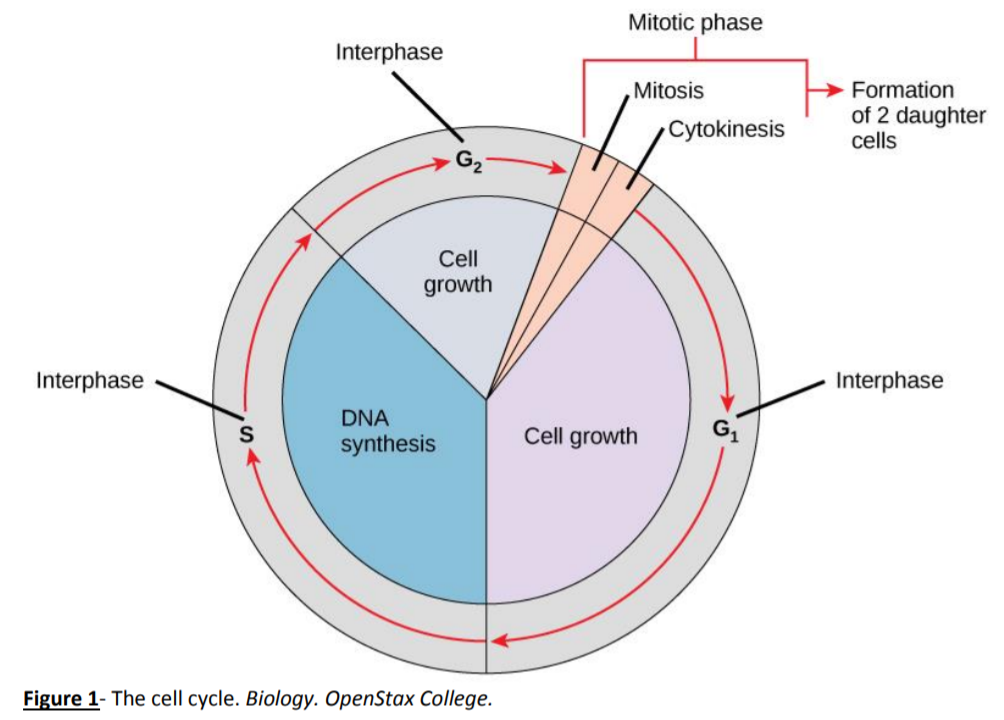
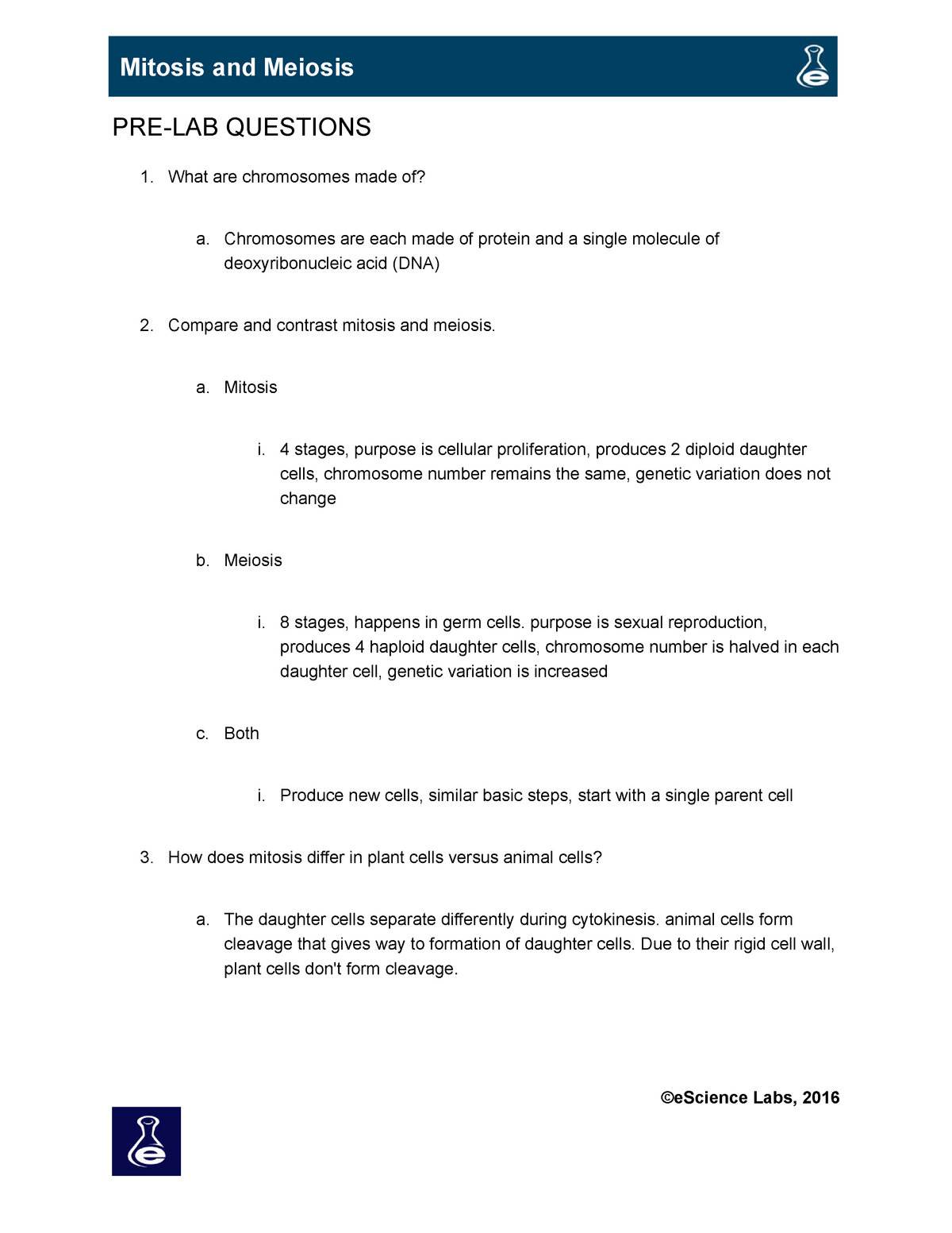
View L12_Meiosis.docx from BSC1005L 1005 at Broward College. Meiosis EXPERIMENT 1: FOLLOWING CHROMOSOMAL DNA MOVEMENT THROUGH MEIOSIS Trial 1 - Meiotic Division Beads Diagram Prophase I Metaphase stages of meiotic division (prophase I and II, metaphase I and II, anaphase I and II, telophase I and II, and cytokinesis). 12. Diagram the corresponding images for each stage in the section titled "Trial 2 Meiotic Division Beads Diagram". Be sure to indicate the number of chromosomes present in each cell for each phase. Also, indicate how ...
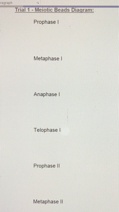
Insert picture here. Part 1 - Meiotic Division Beads Diagram. Prophase I. Metaphase I. Anaphase I.
Meiotic division beads diagram
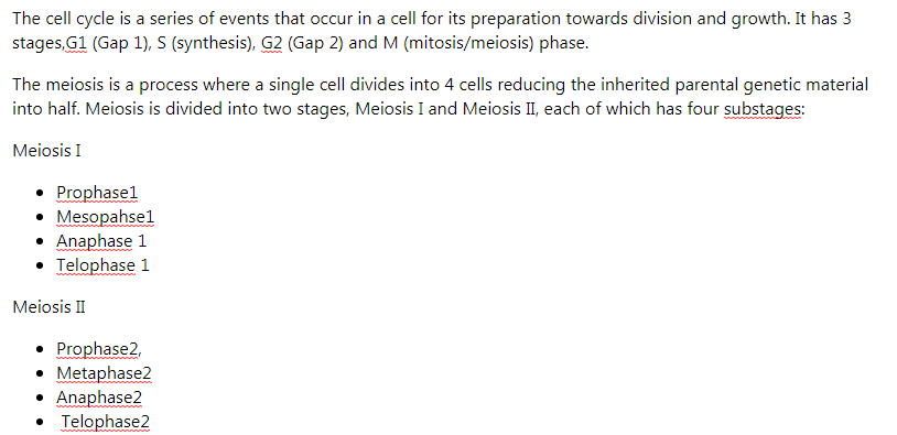
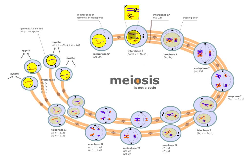
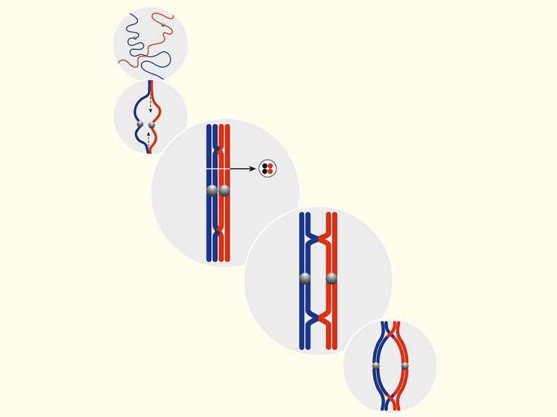
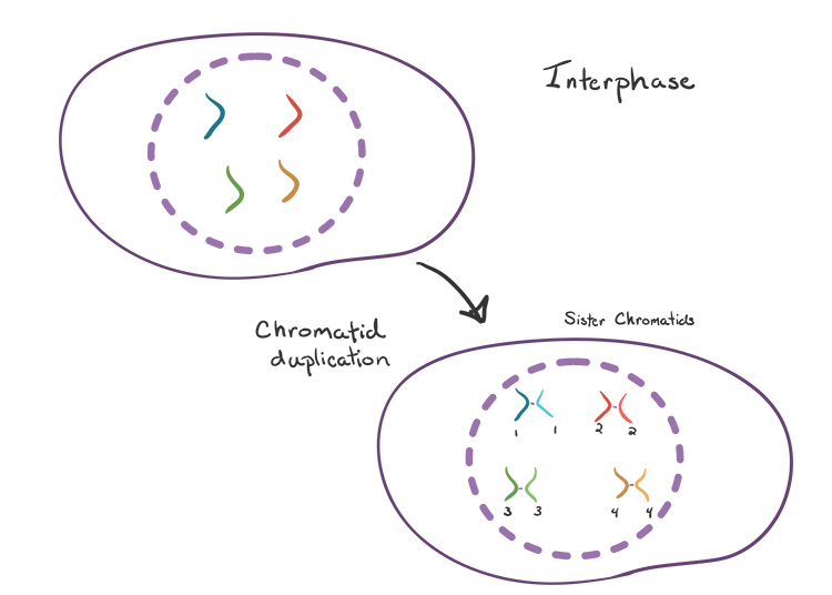
Cell Cycle Division: Part 2 - Meiotic Bead Diagrams (With Crossing Over) Prophase I: One chromosome from mother one from father come. together and wrap around each other so closely that portions of one switch. with portions of the other. They are also lined up along the middle. Anaphase II: The chromosomes are split in half and move to the. side The second meiotic division is where sister (duplicated) chromatids separate. It resembles mitosis of a haploid cell. At the start of the second division, each cell contains 1N chromosomes, each consisting of a pair of sister chromatids joined at the centromere. Here is a simplified diagram illustrating the overall process and products of meiosis: Diagram the corresponding images for each stage in the section titled "Trial 2 Meiotic Division Beads Diagram". Be sure to indicate the number of chromosomes present in each cell for each phase. Also, indicate how the crossing over affected the genetic content in the gametes from Part1 versus Part 2.
Meiotic division beads diagram. Professional academic writers. Our global writing staff includes experienced ENL & ESL academic writers in a variety of disciplines. This lets us find the most appropriate writer for any type of assignment. Nov 15, 2021 · B, Venn diagram showing the overlap between pistil CHH hyper-DMRs (pistils > seedlings) and CHH hypo-DMRs of osrdr2–3 in pistils. Box plots indicate the methylation levels of CHH and 24-nt siRNA abundance on the common and specific regions in pistils or seedlings of the indicated genotypes (** P < 0.01 by two-tailed z-test). Experiment 1: Following Chromosomal DNA Movement through Meiosis Data Tables and Post-Lab Assessment Trial 1 - Meiotic Division Beads Diagram: Biol 103 papers , exams and assignments and many more for students. At Maryland homework we offer assignments and exams from students just like you who have got A grades on these papers. pop beads. In lab, pop beads with magnetic centromeres are used to simulate chromosomes as they move through meiosis. The meiotic board used includes six circles: one on top; two in the middle and four at the bottom. These circles represent the chromosomal make up of the seven nuclei involved in one meiotic event.
Diagram the corresponding images for each stage in the section titled "Trial 2 - Meiotic Division Beads Diagram". Be sure to indicate the number of chromosomes present in each cell for each phase. Also, indicate how the crossing over affected the genetic content in the gametes from Part1 versus Part 2. Part 2 - Meiotic Division Beads ... Diagram the corresponding images for each stage in the sections titled "Trial 1 - Meiotic Division Beads Diagram". Be sure to indicate the number of chromosomes present in each cell for each phase. Disassemble the beads used in Trial 1. You will need to recycle these beads for a second meiosis trial in Steps 7 - 11. Meiotic Division of Cell (With Diagram) In this article we will discuss about the meiotic division of a cell. The meiotic division includes two complete divisions of a diploid cell resulting into four haploid nuclei. The first meiotic division includes a long prophase in which the homologous chromosomes become closely associated to each other ... 5. Diagram the corresponding images for each stage in the sections titled "Trial 1 - Meiotic Division. Beads Diagram". Be sure to indicate the number of chromosomes present in each cell for each phase. 6. Disassemble the beads used in Part 1. You will need to recycle these beads for a second meiosis trial. in Steps 7 - 12.
5. Diagram the corresponding images for each stage in the sections titled "Trial 1 - Meiotic Division Beads Diagram". Be sure to indicate the number of chromosomes present in each cell for each phase. 6. Disassemble the beads used in Part 1. You will need to recycle these beads for a second meiosis trial in Steps 7 - 12. In experiment of Following chromosomal DNA movement through meiosis, what is the trial 1 and trial 2 meiotic division beads diagram for prophase l, metaphase l, anapahse l, telophase l, prophase ll, metaphase ll, anaphase ll, telophase ll, and cytokinesis? Start your trial now! First week only $4.99! arrow_forward. Diagram the corresponding images for each stage in the section titled "Trial 2 - Meiotic Division Beads Diagram". Be sure to indicate the number of chromosomes present in each cell for each phase. Also, indicate how the crossing over affected the genetic content in the gametes from Part1 versus Part 2. stages of meiotic division (prophase I and II, metaphase I and II, anaphase I and II, telophase I and II, and cytokinesis). 12. Diagram the corresponding images for each stage in the section titled "Trial 2 Meiotic Division Beads Diagram". Be sure to indicate the number of chromosomes present in each cell for each phase. Also, indicate how ...
Diagram the corresponding images for each stage in the section titled "Trial 2 - Meiotic Division Beads Diagram". Be sure to indicate the number of chromosomes present in each cell for each phase. Also, indicate how the crossing over affected the genetic content in the gametes from Part1 versus Part 2.
Transcribed image text: Data She experi Lab 12 > Experiment 1 Data Sheet dels TRIAL 2 - MEIOTIC DIVISION BEAD DIAGRAMS: Prophase : LE Metaphase 1: Anaphase 1: Telophase 1: Prophase II: Metaphase II: Anaphase II: Telophase II: Cytokinesis: Data SheeExperia Lab 12 Experiment 1 Data Sheet TRIAL 2 - MEIOTIC DIVISION BEAD DIAGRAMS: Prophase 1 ...

Essential Cell Biology [5 ed.] 0393679535, 9780393679533. Cell biology is taught in classrooms around the world to provide students with a firm conceptual grounding in biology.

5. Diagram the corresponding images for each stage in the sections titled "Trial 1 Meiotic Division Beads Diagram". Be sure to indicate the number of chromosomes present in each phase. 6. Disassemble the beads used in Part 1. You will need to recycle these beads for a second meiosis trial in Steps 8 - 13.
meiotic division (prophase I and II, metaphase I and II, anaphase I and II, telophase I and II, and cytokinesis). Diagram the corresponding images for each stage in the sections titled "Trial 1 - Meiotic Division Beads Diagram". Be sure to indicate the number of chromosomes present in each cell for each phase. Disassemble the beads used ...
6. Diagram the corresponding images for each stage in the sections titled â Trial 1 - Meiotic Division Beads 186 Meiosis Diagramâ . Be sure to indicate the number of chromosomes present in each cell for each phase. 7. Disassemble the beads used in Part 1. You will need to recycle these beads for a second meiosis trial in Steps 8
100% (2 ratings) Meiosis comprises of two phases. In phase one the div …. View the full answer. Transcribed image text: Trial 2 - Meiotic Division Beads Diagram. Previous question Next question.
Biology I Lab Activity - Simulating Mitosis with "Pop Beads" Introduction: Mitosis is the process of one cell dividing to produce two new (daughter) cells (take a look at the diagram . Diagram the corresponding images for each stage in the section titled "Trial 2 - Meiotic Division Beads Diagram".
The latest Lifestyle | Daily Life news, tips, opinion and advice from The Sydney Morning Herald covering life and relationships, beauty, fashion, health & wellbeing

Explore this photo album by Darrietta Lee on Flickr!
Trial 2 - Meiotic Division Beads Diagram: Prophase I. Metaphase I. Anaphase I. Telophase I. Prophase II. Metaphase II. Anaphase II. Telophase I. Cytokinesis. Post-Lab Questions. 1. Poloidy of the DNA at What is the the end of meiosis I? What about at the end of meiosis II?
Trial 2 Meiotic Division Beads Diagram Prophase I 4 Chromosomes Metaphase I 4 from BIOL MISC at Indiana University, Purdue University Indianapolis.

As we have studied, mitosis is the division of the nucleus of somatic (normal body) cells with the intent of making two exact copies of the parent cell. Meiosis ...3 pages
Diagram the corresponding images for each stage in the section titled "Trial 2 - Meiotic Division Bead Diagrams." Be sure to indicate the number of chromosomes present in each cell for each phase. Also, indicate how the crossing over affected the genetic content in the gametes from Part 1 versus Part 2.
Discussion on Meiotic Division Beads Diagram · Start with 20 beads of the same color to create your first sister chromatid pair. · Assemble a second pair of ...
Trial 1 - Meiotic Division Beads Diagram. Part 2: Modeling Meiosis with Crossing Over Part 2 - Meiotic Division Beads Diagram: Image of page 3. Info icon This preview has intentionally blurred sections.Lab 4: Meiosis and Vertebrate Reproduction LAB SYNOPSIS: • Meiosis will be modeled using pop-beads. • The genetic diversity of gametes will ...
Diagram the corresponding images for each stage in the section titled "Trial 2 - Meiotic Division Beads Diagram". Be sure to indicate the number of chromosomes present in each cell for each phase. Also, indicate how the crossing over affected the genetic content in the gametes from Trial 1 versus Trial 2. Trial 2 - Meiotic Division Beads ...
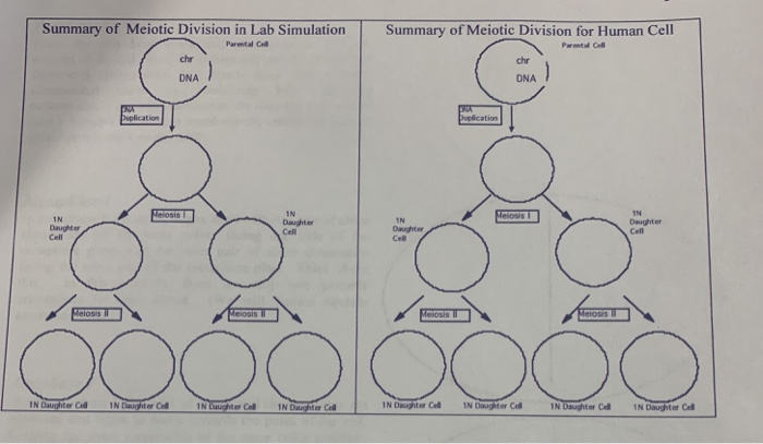
Part 2: Meiotic Division Beads Diagram with Crossing Over. Prophase I Metaphase I Anaphase I Telophase I Prophase II Metaphase II Anaphase II Telophase II Cytokinesis Post-Lab Questions 1. What is the ploidy of the DNA at the end of meiosis I? What about at the end of meiosis II? 2. How are meiosis I and meiosis II different?
May 23, 2020 · 109 Likes, 2 Comments - Dr Raymond C Lee MD (@drrayleemd) on Instagram: “What an amazing virtual aats. Congratulations to my chairman Dr Vaughn Starnes 100th AATS…”

Diagram the corresponding images for each stage in the section titled "Trial 2 - Meiotic Division Beads Diagram". Be sure to indicate the number of chromosomes present in each cell for each phase. Also, indicate how the crossing over affected the genetic content in the gametes from Part1 versus Part 2. Part 2 - Meiotic Division Beads ...
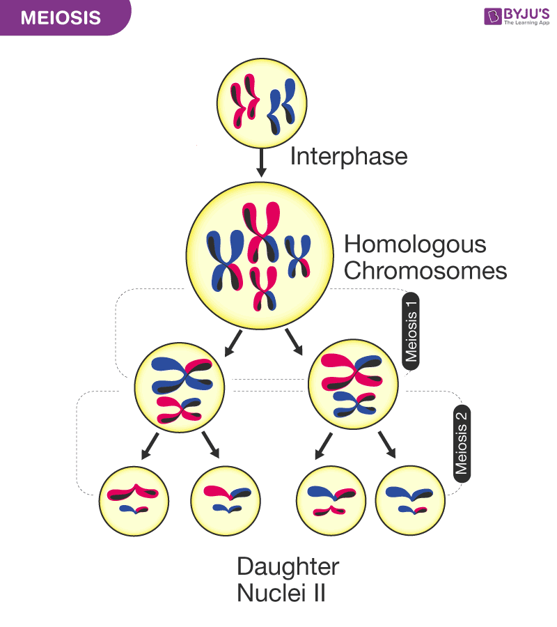
Diagram the corresponding images for each stage in the sections titled "Trial 1 - Meiotic Division Beads Diagram". Be sure to indicate the number of chromosomes present in each cell for each phase. Disassemble the beads used in Part 1. You will need to recycle these beads for a second meiosis trial in Steps 7 - 12.
Diagram the corresponding images for each stage in the section titled "Trial 2 Meiotic Division Beads Diagram". Be sure to indicate the number of chromosomes present in each cell for each phase. Also, indicate how the crossing over affected the genetic content in the gametes from Part1 versus Part 2.

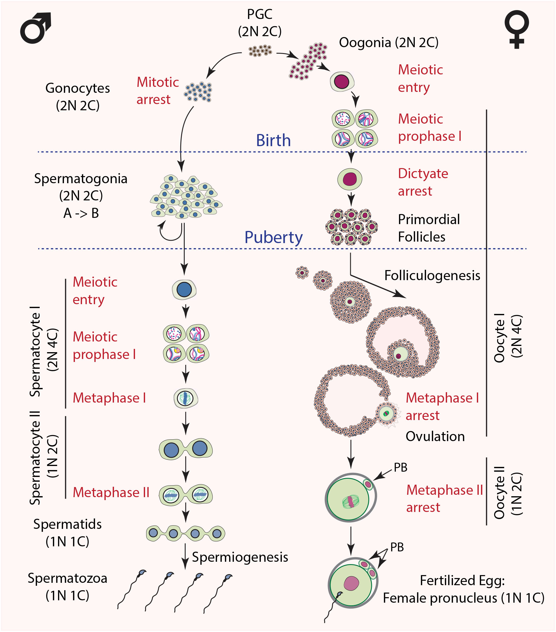
The second meiotic division is where sister (duplicated) chromatids separate. It resembles mitosis of a haploid cell. At the start of the second division, each cell contains 1N chromosomes, each consisting of a pair of sister chromatids joined at the centromere. Here is a simplified diagram illustrating the overall process and products of meiosis:
Cell Cycle Division: Part 2 - Meiotic Bead Diagrams (With Crossing Over) Prophase I: One chromosome from mother one from father come. together and wrap around each other so closely that portions of one switch. with portions of the other. They are also lined up along the middle. Anaphase II: The chromosomes are split in half and move to the. side


0 Response to "38 meiotic division beads diagram"
Post a Comment